
Blog

Historien, nuläget och framtiden för adhesiv tandvård - Intervju med Prof. Bart Van Meerbeek


Som en av huvudredaktörerna för Journal of Adhesive Dentistry, är Prof. Bart Van Meerbeek en av de mest respekterade auktoriteterna inom ämnet dentala bondingmaterial. Här diskuterar han hur de har utvecklats över de tre senaste decennierna och hur framtiden för adhesiv tandvård kan tänkas se ut.


Prof. Van Meerbeek, hur har bondingmaterialen förändrats och utvecklats sedan du först började studera dem?

Jag tror att de stora framstegen den dentala adhesiva teknologin gjort under de senaste 30 åren, och framför allt bondingmaterialens utveckling, har haft en stor påverkan på tandvården i allmänhet och självklart på den restaurativa tandvården i synnerhet. Många av de nuvarande restaurativa dentala procedurerna drar nytta av adhesiva material och tekniker och har avancerat betydligt jämfört med när jag skrev min avhandling för mer än två decennier sedan på ämnet adhesion till dentin. Adhesion till emalj är, naturligtvis, relativt lätt att åstadkomma i jämförelse med adhesion till dentin och, när jag först började forska på detta ämne, var jag begränsad till att utföra kliniska tester där vi konfronterades med ett relativt högt antal förlorade restaurationer på kort tid. Jag har haft turen att på nära håll kunna följa den utveckling dental bonding har genomgått, efter att ha bedrivit forskning inom det här fältet i närmare 30 år nu.


Vid en tidpunkt, började forskningsvärlden inse att det finns ett “smear layer” i mellanskiktet, som uppstår vid preparation av kaviteten, och detta lager inverkar på bindningen. Om du vill uppnå en framgångsrik mikromekanisk och kemisk bindning till substansen, behöver du först göra något åt detta smear layer.


Därefter, kom tiden för conditioners och primers. Tidigare hade de verksamma i den restaurativa vården varit lite rädda för att använda fosforsyra, på grund av dess potential för pulpairritation. Men mer och mer, började tandläkare att använda etsmedel som innehöll denna kemikalie, såväl som primers som effektivt förbättrade bindningen mellan det adhesiva resinet och dentin. Efter att ha uppnått en utmärkt bondingeffekt med flerstegsadhesiver i laboratoriet, vilket senare bekräftades i kliniska studier, fokuserade den fortsatta designen och utvecklingen av adhesiva material på förenkling och förkortning av bondingprocedurerna.


Ur detta kom två typer av adhesiver, som använde två olika metoder för bonding, att utvecklas: etsa-och-skölj-adhesiver och de självetsande, eller etsa-och-torka-adhesiver. Den senaste generationen av universaladhesiver låter nu tandläkaren välja vilken av de två metoderna som ska användas, med samma adhesiv.


Vilka fördelar ger bondade restaurationer jämfört med mer traditionella metoder?

Bondade restaurationer är minimalt invasiva – tandläkaren behöver inte avlägsna frisk vävnad för att skapa underskär för att hålla restaurationen på plats, vilket tillåter en mer konservativ approach. Att behålla så mycket emalj som möjligt ska vara ett mål för alla restaurativa procedurer, eftersom den helt enkelt är den bästa vävnaden att bonda till. Trots att bonding till dentin alltid har varit mer utmanande och faktiskt har bromsat våra strävanden under lång tid, kan adhesiv restauration av tänder, även innefattande effektiv bonding till dentin, numer uppnås på ett pålitligt, förutsägbart och hållbart sätt.


Tillsammans med mycket framgångsrik implantologi för att ersätta saknade tänder, vilket minskar behovet av broar, har restaurationer av enstaka tänder ökat avsevärt i antal. Bonding gynnade ytterligare övergången från vävnadsinvasiva kronor till vävnadsbevarande partiella tandrestaurationer, eftersom moderna adhesiver kan hålla dessa partiella restaurationer på plats på ganska flata och även icke-retentiva ytor. Bondingprocedurer tillåter dessutom att man uppnår restaurationer med ett mer naturligt utseende, genom tekniker att adhesivt cementera estetiska restaurationer av glaskeramik och även starka zirkoniakeramer, som inte ens de längre kan anses vara omöjliga att bonda.


Vad är din åsikt angående den nuvarande generationen av universella adhesivsystem?

Jag tycker att den här generationen adhesiver är mycket bra, men att de fortfarande inte alltid är lika bra som den mer traditionella gyllene standarden med tvåstegs självetsande eller trestegs etsa-och-sköIj-adhesiver vad beträffar den verkliga potentialen för bonding till dental vävnad. Jag ser det dock som positivt att många av dessa universaladhesiver har integrerat MDP-monomeren, vilken bör anses vara en av de mest funktionella monomerer som finns i dag, även om den behöver finnas i hög koncentration och med hög renhet.


MDP-monomeren är, i generall mening, även förträfflig för bonding till zirkonia. När det handlar om bonding till olika typer av keramer såväl som resinbaserade kompositmaterial, är det alltid värdefullt att veta vilka universaladhesiver som innehåller silan och sägs inte längre vara i behov av ytterligare förbehandling av restaurationen. Detta ger fördelarna med lägre teknikkänslighet och färre steg i proceduren – förutsatt att de, faktiskt, fungerar. Det finns nya vetenskapliga bevis för att de silan som ingår i dagens sura vattenbaserade universaladhesiver, dock inte är tillräckligt stabila. Lyckligtvis pågår forskning för att utveckla nya universaladhesiver som innehåller andra silaner med högre stabilitet i vatten och vid högre surhetsgrad.


Framför allt, tror jag att en primer för restaurationer som innehåller en hög koncentration av silan tillsammans med MDP-monomeren fortfarande är mer effektiv än många universaladhesiver för bonding till restaurationsmaterial, eftersom dessa universaladhesiver kan innehålla flera andra ingredienser som skapar en slags konkurrens inom materialet att nå och interagera med substratets yta, vilket leder till sämre bindning.


Ett annat tillkortakommande för universaladhesiver är deras tunna fimtjocklek och relativt höga hydrofilicitet, vilket gynnar vattenupptag och därmed gör dem känsliga för hydrolytisk nedbrytning. I ljuset av detta, är det viktigt att notera att när en viskös och hydrofob flytande komposit appliceras på en universaladhesiv, kan det i viss mån kompensera för detta och tillåta att en hållbar bindning uppstår.


År MDP-monomeren avgörande för den slutliga framgången för universaladhesiver? Finns det andra faktorer som kan påverka detta?

Nåväl, det är väldigt klart att MDP-monomeren är en av de mest effektiva monomererna som finns, tack vare dess primära kemiska bindningspotential till hydroxylapatit. Hur som helst, så finns det signifikanta skillnader på MDP-monomerernas renhet och koncentrationsnivåer i dessa produkter, faktorer som påverkas av om monomeren är syntetiserad av företaget självt, eller om den processen är outsourcad. I grund och botten, en universell adhesiv som innehåller en hög koncentration av mycket ren MDP-monomer borde fungera bäst.


Finns det några specifika fördelar som utmärker självetsande adhesiver?

Den största fördelen är att de inte tar bort all hydroxylapatit och mineraler som finns i dentinet och håller på så sätt det svagare dentinala kollagenet skyddat. Etsning med fosforsyra resulterar i relativt djup och total demineralisering med exponering av kollagenet, vilket gör bindningen mer benägen att degradera. Att delvis behålla mineraler runt kollagenet med användning av en milt självetsande adhesiv tillåter dessutom att att en stark jonbindning uppkommer, i synnerhet när adhesiven innehåller den funktionella monomeren MDP. Man ska dessutom vara medveten om att, även om kemisk bindning inte nödvändigtvis leder till högre bindningsstyrka, kan den skapa bättre hållbarhet på lång sikt.


Vad ser du som nästa steg för den adhesiva tandvården?

En möjlighet är att reducera antalet steg i adhesionsprocessen, med det slutliga målet att ha självadhesiva restaurationsmaterial. Det har skett en utveckling i denna riktning, inklusive studier och kommersiella produkter, men produkterna har inte alltid visat sig vara speciellt effektiva och bindningens hållbarhet har varit oklar. Nu dock, kommer nyare material på marknaden med anspråk på att kunna användas utan någon förbehandling. Deras kliniska effektivitet måste, icke desto mindre, fortfarande bevisas och garanteras innan sådana självadhesiva restaurationsmaterial kan användas som verkliga alternativ i rutinerna för den dagliga praktiken.


En annan möjlighet, och en nuvarande hype inom forskning och utveckling, är utvecklingen av bioaktiva adhesiver. Många dentala forskare och många företag vill att adhesiver inte bara ska ge en god bindning, utan även ha vissa terapeutiska fördelar. Vad exakt en bioaktiv adhesiv egentligen är beror på vem du pratar med. Vissa forskare tycker att de ska ha antibakteriella egenskaper, medan andra konstaterar att remineralisering av dentin och interaktion med pulpaceller krävs för att de ska kvalificera som “bioaktiva”. Vi behöver definitivt undersöka om det är möjligt att vi kan ge dessa material dessa ytterligare egenskaper, men på ett villkor: att de adhesiva materialen inte tappar något av sin ursprungliga bindningsförmåga. Detta är, enligt min åsikt, den största utmaningen för den framtida adhesiva tandvården.
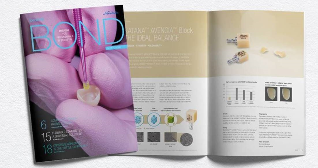
Our latest "BOND" magazine is available now!

Volume 7 of our "BOND" magazine is now published and ready to read!


Content Highlights:

  • Composites versus hybrid ceramics
  • Flowable composites a universal solution?
  • Universal adhesives, is one bottle sufficient?


Start Reading: BOND | VOLUME 7 | 10/2020


Recording 14.10.2020 17:00 CEST - Dr Alessandro Devigus on Dental photography as valuable documentation aid

Dental photography as a valuable documentation aid

The main purpose of photography in dentistry is documentation. The aim is to collect as much information as possible under reproducible conditions. For this purpose, the camera used, its accessories and the format and lighting of the intra- and extra oral recordings relevant to dentistry must be standardized. Under standardized photographic conditions, the recordings made can be compared with one another, even if the later recordings are made only after a long period of time and by different photographers. Only in this form can dental photography be used as a valuable documentation aid. The standards described by Wolfgang Bengel in 1985 are still valid, but they have to be updated and adapted to technical innovations.


In this webinar, the current development in the field of dental photography and related areas will be briefly explained.


Dr. Alessandro Devigus has had his own practice with a focus on CEREC since 1990, editor-in-chief of the International Journal of Esthetic Dentistry.


Topic: Dental photography as a valuable documentation aid
Date: Wednesday. October 14, 2020 - 17:00 - 18:30 CEST
Time: approx. 90 min (including discussion and questions)


Recording 16.09.2020 17.00 CEST - Dr Dirawi on Zirconia; What's new with this old material?

ZIRCONIA: NEW INSIGHTS INTO TRIED AND PROVEN MATERIAL

You learn from your mistakes, but it’s better if you can learn from those of others. Dr. Dirawi will share his experience, tips and tricks when it comes to creating perfect Zirconia restorations.
Having great material like KATANA Zirconia is an excellent starting point. Yet to achieve highly aesthetic outcome you need to be aware of the unexpected pitfalls and challenges that can be prevented.
Join our webinar and learn what Dr. Dirawi has to say after years of using Zirconia. Bonus – we will have a conversation about cementation of zirconia as well, to achieve durable robust results.


DR WISSAM DIRAWI DDS, MALMÖ, SWEDEN

  • Teacher at Malmö Dental University in Sweden
  • Researcher in the fields of ceramics and implant complications
  • Long clinical experience from public dental care and private dental care
  • Expert in ceramics, implants and digital dentistry
  • 2018 - Specialist in Oral Prosthodontics
  • 2000 - DDS


Clinical case with CLEARFIL MAJESTY™ Posterior

By Magdalena Osiewicz, DDS, MSc, PhD


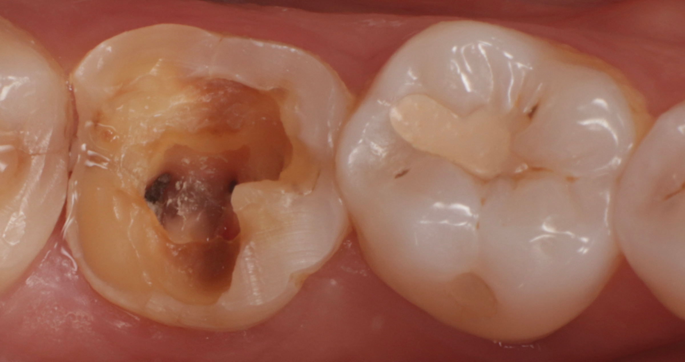
Fig. 1 Defective composite resin restoration in molars.


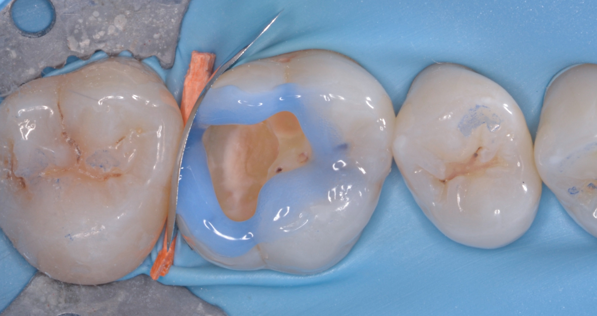
Fig. 2 Application of CLEARFIL™ SE BOND 2 to cavities.


Fig. 3 Restoration of the cavities with CLEARFIL MAJESTY™ Posterior in the A2 Classic shade.


Fig. 4 Final restorations of Class I and II with CLEARFIL MAJESTY™ Posterior and polish with CLEARFIL™ Twist DIA.


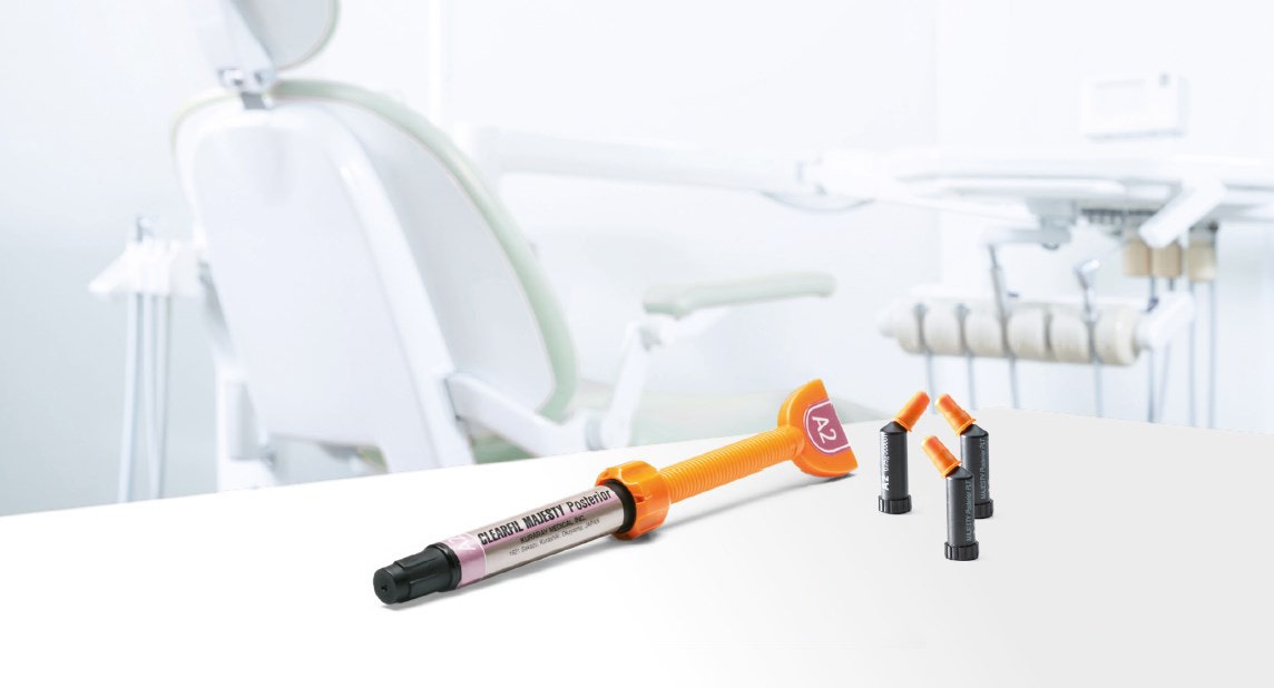
CLEARFIL MAJESTY™ Posterior is a resin composite with high strength and great optical properties developed for posterior restorations and suitable even for the most demanding patients. Figure 1 shows the initial clinical situation with insufficient resin composite restorations in the lower molars. After removal of the old fillings, the cavities were treated with CLEARFIL™ SE BOND 2 (Figure 2).


Then, I restored them with CLEARFIL MAJESTY™ Posterior in the A2 Classic shade (Figure 3). The fissures were highlighted with brown color modifier. Finally, finishing was performed in three steps: The excess of composite resin was removed with a fine-grained diamond bur. Final contouring was accomplished with a carbide bur, before CLEARFIL™ Twist DIA was used to obtain a natural gloss (Figure 4).


CLEARFIL MAJESTY™ Posterior is characterized by high mechanical strength, hardness and bending strength, a low coefficient of thermal expansion, low polymerization shrinkage and good aesthetics. Due to these features and a reliable long-term behavior, CLEARFIL MAJESTY™ Posterior should have a place in every dental office for direct posterior restorations. Excellent outcomes are achievable and therefore I recommend its use.


Dentist:

Magdalena Osiewicz, DDS, MSc, PhD


Department of Integrated Dentistry, Jagiellonian University, Krakow, Poland.

Department of Dental Materials Science, Academic Centre for Dentistry Amsterdam (ACTA), University of Amsterdam and Vrije Universiteit Amsterdam, Amsterdam, the Netherlands.

Department of Integrated Dentistry, Jagiellonian University, Krakow, Poland.

Department of Dental Materials Science, Academic Centre for Dentistry Amsterdam (ACTA), University of Amsterdam and Vrije Universiteit Amsterdam, Amsterdam, the Netherlands.


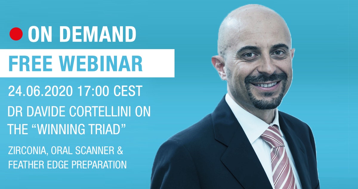
Recording 24.06.2020 17.00 CEST - Dr Davide Cortellini on the “winning triad”

Zirconia, Oral scanner, feather edge preparation: the “winning triad”

The introduction of digital technology associated with the development of the latest zirconia materials offers the clinical and the dental technician a wide range of solutions to achieve aesthetics and function in a simple, predictable and above all conservative way. A further type of vertical preparation called "Minimally Invasive Vertical Preparation" has been introduced, applicable to numerous prosthetic clinical situations including cases of aesthetics. Minimally invasive vertical preparations, new type of zirconia and intraoral scanners represent a “winner triad” for today's prosthetic therapy.


ABOUT DR DAVIDE CORTELLINI

Graduated with honors in Dentistry at the University of Siena in 1992, wins the scholarship of the Italian Society of Periodontology (SIDP) for the year 1994-95. He therefore has the opportunity to attend the Department of Periodontology and Fixed Prosthesis of Prof. NP. Lang at the University of Bern in Switzerland, carrying out clinical and research activities. He obtained the title of "Doctor Medicinae Dentium" (D.M.D.) at the same University in 2000.

Author of scientific publications in international journals, he is an Active Member  of the Italian Academy of Prosthetic Dentistry (AIOP). Active member of the International Academy of Digital Dentistry (IADDM). He lectures in Italy and abroad, and he owns his private practice in Riccione mainly oriented to digital, aesthetic and complex prosthetic rehabilitations.


EC Certificate of Kuraray Noritake Dental Inc. products

At Kuraray Noritake Dental Inc. we are proud that our products are being developed using innovative technologies, paying attention to detail and keeping in mind the needs of our customers.


We are committed to offering outstanding products that provide dental professionals with the flexibility for self-expression and creativity, adding value and delivering superb aesthetic results to each and every procedure.


Usability, simplicity and, of course, quality always in mind - providing dental professionals with materials that will make their daily practice stress free.


As a prove that our products meet the needs of dentists and lab technicians alike, we are happy to share with you the EC Certificates of Full Quality Assurance System that we have received from The Notified Body.  Those can be downloaded below.


Chairside products


Labside products


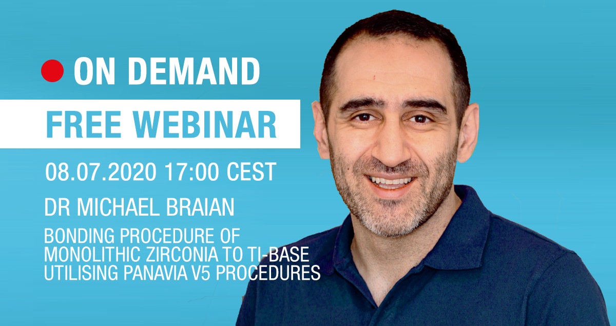
Recording 08.07.2020 17.00 CEST - Dr Michael Braian on the bonding procedure of monolithic Zirconia to Ti-base utilising PANAVIA V5 procedures

Join Dr Michael Braian on the webinar focusing on the bonding procedure of monolithic Zirconia to Ti-base utilising PANAVIA V5 procedures. 


ABOUT DR MICHAEL BRAIAN

Michael Braian, DDS, CDT, PhD is an international reference in the Digital Dentistry area as he has been working with technology in the dental field for almost 15 years. His professional profile combines every key aspect in dentistry as he is a Dentist, Certified Dental Technician and has a PhD in Digital Dentistry by Malmö University.


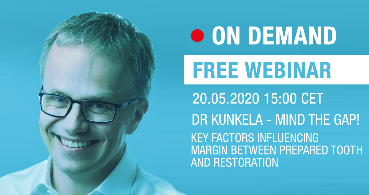
Recording - 20.05.2020 15.00 CET - Dr Josef Kunkela on Mind the Gap!

Mind the Gap!

Webinar about the key factors which influence the gap between the margin of prepared tooth and restoration, also about accuracy evaluations of milled materials and resin cement removal techniques during cementation.


Dr JOSEF KUNKELA, DMD, PHD

1993
Dentistry graduate of First Medical Faculty of Charles University, Prague, Czech Republic

1995
Fellow in Medical Faculty of Charles University Prosthodontic Department in Hradec Králové, Czech Republic

1999
Gained Second Degree Specialization in Prosthodontics

2001
Named external teacher of the Prosthodontic Department at the Charles University Clinic of Dentistry in Hradec Králové and Palacky University in Olomouc, Czech republic

2009
Named president of Czech Society of CAD/CAM Dentistry

2010
Founder of KUNKELA Campus, International CAD/CAM Traning Center in Jindrichuv Hradec, Czech Republic (Certified Exclusive DentsplySirona Training Center)

2018
Finished postgraduate doctoral degree PhD, thesis Dental Office Management and Marketing (University of Economics Prague)


Certified CEREC trainer by the International Society of Computerized Dentistry (ISCD)

Member of DSD (Digital Smile Design) Master Team

Member of MicroVision Group

Member of SKYN Concept Team

Member of Academy of Digital Dentistry

Board Member of ADDA (Association of Digital Dentistry Academies)

Ambassador of DDS (Digital Dentistry Society)

CEREC Beta Tester

CEREC Guide Beta Tester

CEREC Advocate

Key Opinion Leader in the area of digital dentistry

International speaker at the field of Digital Dentistry, Guided Implantology, Management & Marketing

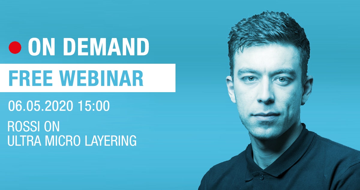
Recording - 06.05.2020 15.00 - Roberto Rossi on Ultra Microlayering

New liquid ceramic FC Paste Stain for full-contour solutions with KATANA Multi-layered Zirconia - Features and 3D-technique.


ROBERTO ROSSI 

"YOU CAN’T STOP THE WAVES, BUT YOU CAN LEARN TO SURF"

Born in Savona in 1989, he studied and got his diploma at “Mazzini” Dental School, the one dental school in his hometown, and he still lives there. In 2007 he was awarded the National Award for Best Dental Technician in Turin. Since 2008 he has been working at Daniele Rondoni’s Dental Lab and he is now in charge of the aesthetic planning of dental restorations. He shares this task with Master Dental Technician Daniele Rondoni, with whom he decides which strategy and materials – composites or ceramic especially – to opt for. In 2011 he coauthored with Mr. Rondoni “Sei faccette additive in composito” (Six additional composite facets), an article published in Dental Labor, 5/2011. A teacher at NISC, Noritake Italian Study Club since 2014, he is also a teacher at the AAT Community College – a reality he feels especially attached to – and he is in charge of the photographic services and social network profiling of the lab.

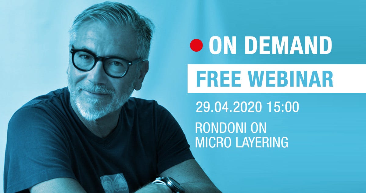
Recording 29.04.2020 15.00 - Daniele Rondoni - "When art meets Technology"

When art meets technology;
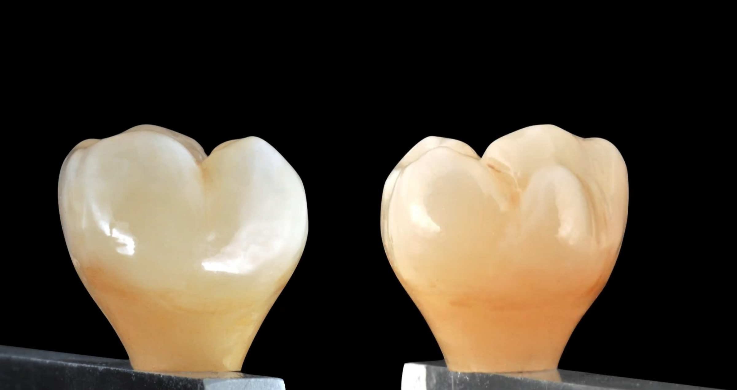
Logical evolution of design and techniques: Microlayering with Kuraray Noritake new porcelains. 


DANIEL RONDONI
Born in Savona in 1961, he lives and works in his hometown where he has been the manager and director of his own laboratory since 1982.

He got his Dental Technician Degree at "P. Gaslini" Professional Institute in Genoa in 1979 and in 1981  was one of the professionals who started the Dental Technician School in Savona as a teacher and a member of the founding Council.

His career features numerous international professional experiences in Switzerland, Germany and Japan and since 2007 he has been accepted as an active member of the EAED.

In 1994 he started an international lecturing career in many of the most prestigious dental symposiums around the world.Particularly devoted to the study of morphology and dental aesthetics, he actively collaborates to the development of materials used for aesthetic dental restoration.He authored the text "Tecnica della Multistratificazione in ceramica" (Ceramic Multilayering Technique) and a lab manual about the use of composite materials, aimed at establishing working protocols for both indirect technique and composite pressing on metal structures and implants and thus introducing his own method, named "Sistema di stratificazione a durezza inversa" TENDER (Inverted Hardness Layering System).


- EAED Active Member

- IAED Active Member

- Styleitaliano Honorary member

- SICED Associate and Speaker

- Noritake Dental Materials International Instructor


Clinical case with direct composite applications in anterior teeth

By Dr. PhD. Jusuf Lukarcanin


Is it possible to fulfil high aesthetic demands by restoring anterior teeth with composite resin? It is – provided that several important factors are respected. One of these factors is the faithful reproduction of the natural tooth morphology, which has a decisive impact on aesthetics and function. Moreover, success is determined by the selection of the right shades of high-quality composite resin and their purposeful combination using proper layering techniques.


Introduction

The aesthetic appearance of direct anterior restorations is affected by proper shade selection on the one hand and the creation of a natural shape and texture on the other1. Hence, the dental practitioner’s own artistic skills play a decisive role. According to Fahl, information about the tooth morphology and function, and the optical properties of the tooth should be taken into consideration when the most suitable restorative material and shade are selected2.


These minimally invasive composite restorations are no longer a temporary solution for the anterior region. Instead, they are regarded as an adequate alternative to indirect restorations, as they are both durable and able to closely imitate the natural tooth structure34.


Clinical case example 1

This 45-year-old female patient presented with a diastema and a disproportion in the size and shape of her maxillary central incisors (Fig. 1). In the first step, a detailed case history was taken and an intra-oral examination was carried out. Subsequently, the initial situation was recorded by taking intra-oral photographs, which would allow for a computer-aided morphological evaluation and treatment planning (Fig. 2).

Fig. 1: Pre-operative image.

Fig. 2: Digital mock-up.

The patient’s second visit started with a professional tooth cleaning procedure followed by isolation of the maxillary anterior teeth. Afterwards, the tooth shade was determined and appropriate composite shades were selected. In this case, the shades A2E, Amber Translucent and A3D of CLEARFIL™ Majesty ES-2 Premium (Kuraray Noritake Dental, Japan) appeared to be most suitable. In addition, a mock-up was created using mock-up resin in order to produce a silicone key.


Opting for a minimally invasive procedure, no mechanical tooth preparation using drills was performed after removal of the mock-up. Instead, the enamel was merely etched with 35% phosphoric acid gel (K-Etchant, Kuraray Noritake Dental) to increase the surface roughness. After rinsing and drying, the adhesive agent (CLEARFIL™ Universal Bond, Kuraray Noritake Dental) was applied to the etched surfaces. Composite layering started with the build-up of palatal shells with the aid of the silicone key. Following light-curing of the shells, a small amount of composite in the dentin shade A3D was applied to the proximal surfaces using a thin spatula and a brush. The aim was to reduce light transmission in the area of the dentin core. The restoration was completed with a combination of the composite shades A2E (enamel) and Amber Translucent, which were applied using a modeling brush.


Finishing and polishing was accomplished using flexible rubber polishing discs containing diamond particles (CLEARFIL™ Twist DIA, Kuraray Noritake Dental) with a low-speed handpiece. No additional finishing and contouring was necessary due to the use of a brush during layering, which ensured the creation of a natural shape and surface texture. Figure 3 shows the outcome of the restoration procedure.


Fig. 3: Treatment outcome immediately after polishing.

Oral hygiene training was provided and follow-up examinations were performed after three, six and twelve months (Fig. 4). Healthy hard and soft tissue conditions were observed during these visits.

Fig. 4: Clinical situation at the one-year recall.

Clinical case example 2

This 30-year-old female patient had a diastema, irregularly shaped anterior teeth and showed signs of abrasive tooth wear (Fig. 5). Following a detailed anamnesis and intra-oral examination, the tooth shade was determined and the composite CLEARFIL™ Majesty ES-2 Premium selected in the monochromatic shade Universal A1.

Fig. 5: Pre-operative clinical situation.

Following the isolation of the working field, 35% phosphoric acid etchant (K-Etchant) was applied to the enamel of all teeth between the maxillary right canine and the maxillary left first molar. The surfaces were then treated with a universal bonding agent (CLEARFIL™ Universal Bond) as recommended by the manufacturer. Modeling was carried out with a thin spatula and a modeling brush for composite. Neither a silicone key nor any wetting or modeling resin were used in the procedure. For polishing, the flexible polishing discs CLEARFIL™ Twist DIA were used at low rotational speed. Thanks to the use of the modeling brush, no additional finishing with diamond-coated instruments was necessary. Figures 6 and 7 show the final restoration at baseline and one week after completion of the treatment.


Fig. 6: Treatment outcome at the day of the restorative procedure.


Fig. 7: Clinical situation after one week.

This patient also received oral hygiene training and presented for recalls three, six and twelve months after the treatment. The patient maintained an exemplary oral hygiene behaviour, so that it came as no surprise that the soft tissues were healthy and the restorations were in a perfect condition after one year (Fig. 8).


Fig. 8: Clinical situation one year after the restorative treatment.


Discussion

Nowadays, direct composite restorations are becoming increasingly popular. Especially for young patients and all those who do not want to sacrifice large amounts of healthy tooth structure, the technique is an ideal treatment option5. In many cases, aesthetic outcomes are possible without mechanical tooth preparation, but a selective etching procedure only6.


The clinical lifetime of these restorations depends on many factors. Important prerequisites for high-quality outcomes include the selection of a suitable composite material with the required surface hardness, appropriate finishing and polishing, a good oral hygiene behaviour, and proper maintenance measures during periodical follow-up visits. As a matter of course, the manual skills of the dental practitioner and the use of selected materials according to the manufacturer’s instructions for use also have a direct impact on the long-term success of the restorations789. A user’s inability to meet one of these requirements and failure to carry out all working steps correctly may have a direct impact on the quality of the restoration.


Conclusion

Composite resin is a popular material class for the production of aesthetic anterior restorations die to their straightforward use and rapid application, good repair options and high aesthetic potential when used properly . The two case examples illustrate that a treatment with composite resin is often the best treatment option when a non-invasive procedure completed within a single visit is desired.


About the author

Dr. Jusuf Lukarcanin is a Certified Dental Technician (DCT) and a Doctor of Dental Science (DDS). He studied dentistry at the Ege University Dental Faculty in Izmir, Turkey, where he obtained a Master's degree in 2011. In 2018, he received a PhD degree from the Department of Restorative Dentistry of the same university, where he also worked as a researcher for six years. Between 2012 and 2019, Dr. Lukarcanin was the head doctor and general manager at Ozel Gen Dental Clinic (OZEL GEN Ağız ve Diş Sağlığı Polikliniği) in Izmir. Currently, he works at Tinaztepe GALEN Hospital in the Tinaztepe Health Group of Tinaztepe University and in the Department of Research & Development of GULSA Company in Izmir.


References

1. Heymann HO (1987) The artistry of conservative esthetic dentistry Journal of the American Dental Association 115(Supplement)14-23.

2. Fahl N Jr (2012) Single-shaded direct anterior composite restorations: A simplified technique for enhanced results Compendium of Continuing Education in Dentistry 33(2) 150-154.

3. Barrantes, J. C. R., Araujo Jr, E., & Baratieri, L. N. (2014). Clinical Evaluation of Direct Composite Resin Restorations in Fractured Anterior Teeth. Odovtos-International Journal of Dental Sciences, (16), 47-61.

4. Vargas M (2011) Clinical techniques: Monocromatic vs. polycromatic layering: How to select the appropriate technique ADA Professional Product Review 6(4) 16-17.

5. Ferracane, J. L. (2011). Resin composite—state of the art. Dental materials, 27(1), 29-38.

6. Norling, N. A. (2010). Combining “prep-less” and conservatively prepared veneers to correct enamel defects and asymmetry. Journal of Cosmetic Dentistry, 2010.

7. Ölmez, A., & Kisbet, S. (2012). Kompozit rezin restorasyonlarda bitirme ve polisaj işlemlerindeki yeni gelişmeler. Acta Odontologica Turcica, 30(2), 115-22.

8. Senawongse, P., & Pongprueksa, P. (2007). Surface roughness of nanofill and nanohybrid resin composites after polishing and brushing. Journal of Esthetic and Restorative Dentistry, 19(5), 265-273.

9. Giacomelli, L., Derchi, G., Frustaci, A., Bruno, O., Covani, U., Barone, A., Chiappelli, F. (2010). Surface roughness of commercial composites after different polishing protocols: an analysis with atomic force microscopy. The open dentistry journal, 4, 191.

10. Hickel, R., Heidemann, D., Staehle, H. J., Minnig, P., & Wilson, N. H. F. (2004). Direct composite restorations. Clin Oral Invest, 8, 43-44.

11. Korkut, B., Yanıkoğlu, F., & Günday, M. (2013). Direct composite laminate veneers: three case reports. Journal of dental research, dental clinics, dental prospects, 7(2), 105.

Anmäl dig för nyheter
Gå med tusentals tandläkare och få gratis råd som kan hjälpa dig och din karriär. Vi kommer inte skräppost eller dela din e-post.