Tagged with 'Kuraray Noritake'

10 years KATANA™ Zirconia multi-layered series

Photo credits to Giuliano Moustakis


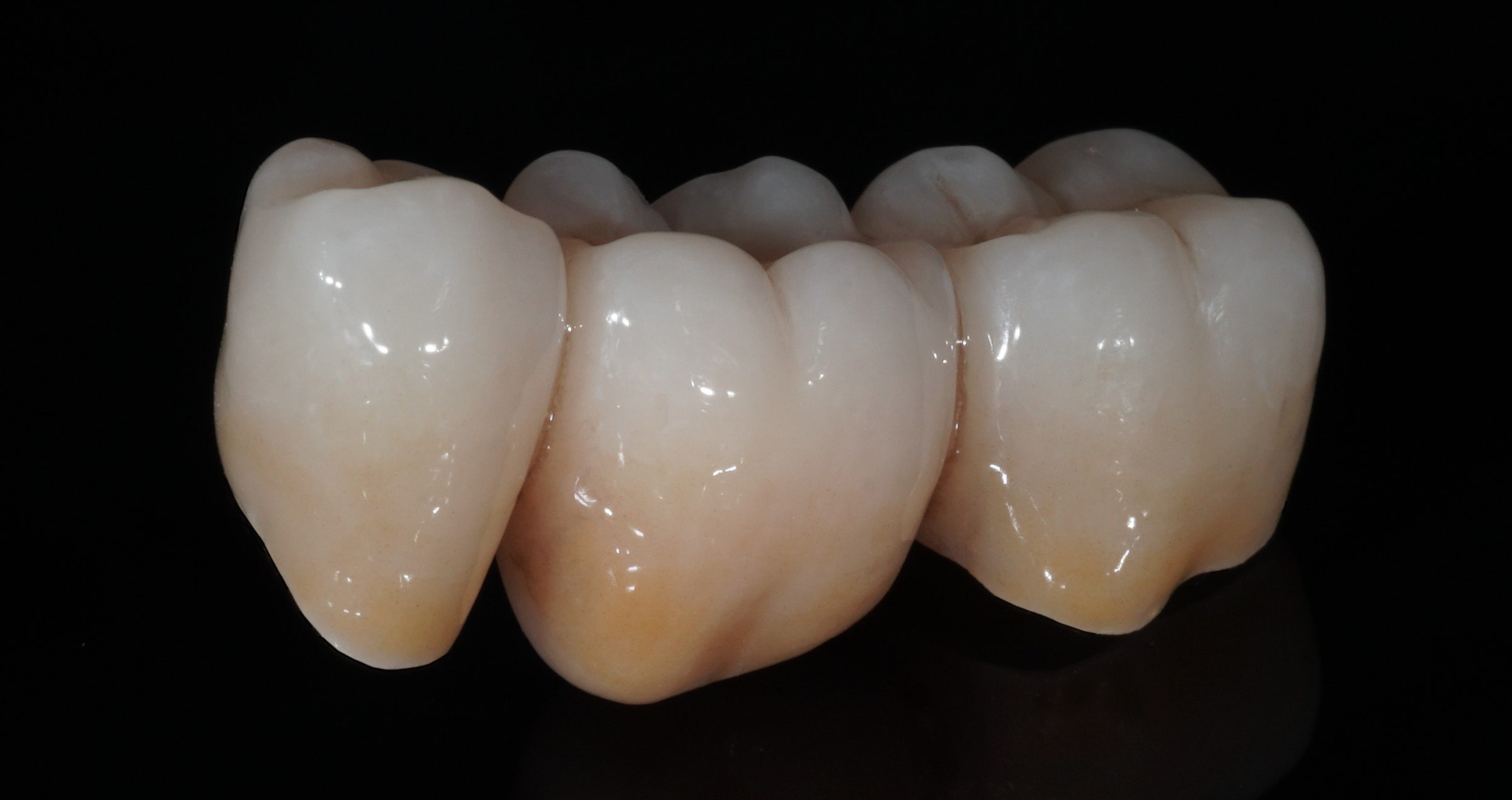
Can you imagine a world without multi-layered zirconia? The invention of a zirconia material with natural colour gradation and well-balanced translucency and strength led to fundamental changes in the way zirconia-based restorations are produced. When the first product of its kind – KATANA™ Zirconia ML – was introduced to the dental market exactly ten years ago, dental technicians all over the world suddenly started rethinking their manufacturing concepts.


Since then, the trend towards a decreased thickness of the porcelain layer, a limiting of this layer to the vestibular area and the production of monolithic restorations is clearly perceivable. Technicians have developed their own concepts of micro-layering, which allow for more patient-centred approaches. This is also due to the fact that the total wall thicknesses of the restorations may be decreased without compromising the aesthetics. The line-up of multi-layered zirconia currently available from Kuraray Noritake Dental Inc. (KATANA™ Zirconia UTML, STML, HTML Plus and KATANA™ Zirconia YML with additional translucency and strength gradation) enables users to make indication-related material choices for the production of restorations that are precisely aligned to the individual demands of each case.


The reasons to choose KATANA™ quality


But why choose KATANA™ Zirconia instead of any other multi-layered zirconia disc? According to experienced users of the KATANA™ Zirconia Multi-Layered series, there are many reasons to opt for KATANA™.


For Jean Chiha, owner at North Star Dental Laboratories and Milling Center in Santa Ana, California, it is the combination of optical and mechanical properties that makes the difference: 


“KATANA™ Zirconia is the game changing material with well-balanced esthetics and strength!”. 


Naoki Hayashi, president of Ultimate Styles Dental Laboratory in Irvine, California, aesthetics is the most decisive argument to opt for the discs from Kuraray Noritake Dental Inc. He states:


“KATANA™ Zirconia discs offer trusted esthetics which gives me confidence in my clinical cases”.


Naoto Yuasa, chief ceramist at Otani Dental Clinic in Tokyo, adds predictability as an important factor:


“KATANA™ sustains my passions for aesthetic restorations and those of a predictable future in the long run”
, whereas dependability is the key element.


For Hiroki Goto, the laboratory manager at Sheets and Paquette Dental Practice in Newport Beach, California reports:


“Without KATANA™ there is no pride. Haven’t experienced it yet? You have to see how reliable it is!”


Finally, we have asked Kazunobu Yamada, a pioneer in making porcelain laminate veneers using complementary color techniques and a first-hour user of KATANA™ Zirconia, what comes to his mind when thinking about KATANA™ Zirconia. According to the president of CUSP Dental Laboratory in Nagoya City, there is a clear link between the product name and its characteristics:

“Did you know that the word "KATANA" also means "protection against misfortune and evil"? Katana zirconia, the culmination of many years of research and development by Kuraray Noritake, has an unparalleled quality. KATANA™, for me, is truly "Protection for all technicians".”


The origin of well-balanced properties


It seems that the KATANA™ Zirconia Multi-Layered line-up stands out due to set of valuable properties enabling a dental technician to produce beautiful, high-quality restorations every time. The secret of success lies in meticulous raw material selection and controlled processing from the powder to the pre- sintered blank carried out at the production facilities of Kuraray Noritake Dental Inc. in Japan. They provide for the high product quality that is responsible for the materials’ outstanding behaviour supporting the best possible outcomes.


Unilateral bite elevation with a zirconia bridge and a lithium disilicate onlay

Clinical case by Dr. Florian Zwiener


The 85-year-old female patient presented after osteosynthesis of a multiple mandibular fracture she had sustained after a fall. During fixation, a massive nonocclusion had occurred in the left posterior region of the mandible (teeth 34 to 37; FDI notation). The patient desired to be able to chew properly again in this area. After endodontic treatment of the two avulsed central incisors, which had been replanted in the hospital, and periodontal therapy, a bite elevation was planned on the left side.


The idea was to restore the teeth and elevate the bite with three onlays and a crown made of lithium disilicate (IPS e.max CAD, Ivoclar Vivadent). During tooth preparation, however, a longitudinal root fracture was detected on the first molar. Therefore, only the first premolar was restored in this session. For this purpose, an onlay was produced chairside (with the CEREC system, Dentsply Sirona) and adhesively luted with PANAVIA™ V5 (Kuraray Noritake Dental Inc.). The first molar was extracted. One week later, the extraction socket, which was still healing, was modelled for the ovoid pontic using an electrotome loop. The second premolar and molar were prepared as abutment teeth for a bridge. The bridge was then milled from KATANA™ Zirconia Block for Bridge in the shade A3.5 and individualized with CERABIEN™ ZR FC Paste Stain (both Kuraray Noritake Dental inc.). After another week, the bridge was luted with the self-adhesive resin cement PANAVIA™ SA Cement Universal (Kuraray Noritake Dental Inc.) following sandblasting.


Fig. 1. Situation after multiple mandibular fracture on the left side.


Fig. 2. Clinical situation at the initial appointment in the dental practice.


Fig. 3. Open bite in the mandibular left posterior region.


Fig. 4. Bridge design …


Fig. 5. … using the CEREC Software.


Fig. 6. Due to the bright shade of the teeth in the cusp area, the restoration was positioned high in the KATANA™ Zirconia Multi-Layered Block.


Fig. 7. Surface texturing in the pre-sintered state (prior to the final sintering procedure).


Fig. 8. Bridge after a seven-hour sintering cycle.


Fig. 9. Appearance of the bridge after individualization with CERABIEN™ ZR FC Paste Stain …


Fig. 10. … and two glaze firings.


Fig. 11. Clinical situation after restoring the teeth with a lithium disilicate onlay and a zirconia bridge.


FINAL SITUATION


Fig. 12. Onlay and bridge in place (after adhesive luting with PANAVIA™ V5 and self-adhesive luting with PANAVIA™ SA Cement Universal).


Fig. 13. Final X-ray used to check for excess cement around the bridge.


Dentist:

DR. FLORIAN ZWIENER


Dr. Florian Zwiener is a distinguished dental professional known for his expertise in Endodontics, Prosthodontics, and CAD/CAM technology. Born in Cologne, Germany, he developed a passion for dentistry and pursued his education at the University of Cologne, where he obtained his degree in Dentistry. Currently, Dr. Florian Zwiener practices at the Dr. Frank Döring Dental Clinic in Hilden, Germany. Here, he continues to apply his specialized knowledge and skills, ensuring that his patients receive the highest quality of care. Follow Dr. Zwiener on Instagram: @dr.florian_zwiener.


Universal adhesives: rationalizing clinical procedures

Case report with Dr. José Ignacio Zorzin


Rationalizing clinical workflows: This is the main reason for the use of universal products in adhesive dentistry. They are suitable for a wide range of indications and different application techniques, fulfil their tasks with fewer components than conventional systems and often involve fewer steps in the clinical procedure. Universal adhesives are a prominent example.


How do universal adhesives contribute to a streamlining of workflows?


When restoring teeth with resin composite, the restorative material will undergo volumetric shrinkage upon curing. By bonding the restorative to the tooth structure with an adhesive, the negative consequences of this shrinkage – marginal gap formation, marginal leakage and staining, hypersensitivity issues and the development of secondary caries – are prevented. The first bonding systems available on the dental market were etch-and-rinse adhesives, which typically consisted of three components: an acid etchant, a primer and a separate adhesive. Later generations combined the primer and the adhesive in one bottle, or were two or one-bottle self-etch adhesives. Universal adhesives (also referred to as multi-mode adhesives) may be used with or without a separate phosphoric acid etchant.


Fig. 1. Volumetric shrinkage of resin composite restoratives and its clinical consequences.


Which technique to choose depends on the indication and the clinical situation. In most cases, the best outcomes are obtained after selective etching of the enamel1. Bonding to enamel is generally found more effective when the enamel is etched with phosphoric acid, while the application of phosphoric acid on large areas of dentin involves the risk of etching deeper than the adhesive is able to hybridize. When the cavity is small, however, selective application of the phosphoric acid etchant to the enamel surface may not be possible, so that a total-etch approach is most appropriate. Finally, in the context of repair, the self-etch approach may be the first choice, as phosphoric acid might impair the bond strength of certain restorative materials by blocking the binding sites. By using a universal adhesive, all these cases may be treated appropriately, as the best suitable etching technique can be selected in every situation.


Apart from the differences related to the use or non-use of phosphoric acid etchant on the enamel or enamel-and-dentin bonding surface, the clinical procedure is always similar with the same universal adhesive. The following clinical case is used to illustrate how to proceed with CLEARFIL™ Universal Bond Quick (Kuraray Noritake Dental Inc.) in the selective enamel etch mode, and it includes some details about the underlying mechanism of adhesion.


How to proceed with selective enamel etching?

A clinical example.


This patient presented with a fractured maxillary lateral incisor, luckily bringing the fragment with him. Hence, it was decided to adhesively lute the fragment to the tooth with an aesthetic flowable resin composite.


Fig. 2. Patient with a fractured maxillary lateral incisor.


Fig. 3. Close-up of the fractured tooth.


Fig. 4. Working field isolated with rubber dam.


As proper isolation of the working field makes the dental practitioner’s life easier, a rubber dam was placed using the split-dam technique. It works well in the anterior region of the maxilla, as the risk of contamination with saliva from the palate is minimal. Once the rubber dam was placed, the bonding surfaces needed to be slightly roughened to refresh the dentin. As the surfaces were also slightly contaminated with blood and it is important to have a completely clean surface for bonding, KATANA™ Cleaner was subsequently applied to the tooth structure, rubbed into the surfaces for ten seconds and then rinsed off. The cleaning agent contains MDP salt with surface-active characteristics that remove all the organic substances from the substrate. The fragment was fixed on a ball-shaped plugger with (polymerised) composite and also cleaned with KATANA™ Cleaner.


Fig. 5. Cleaning of the tooth …


Fig. 6. … and the fragment with KATANA™ Cleaner.


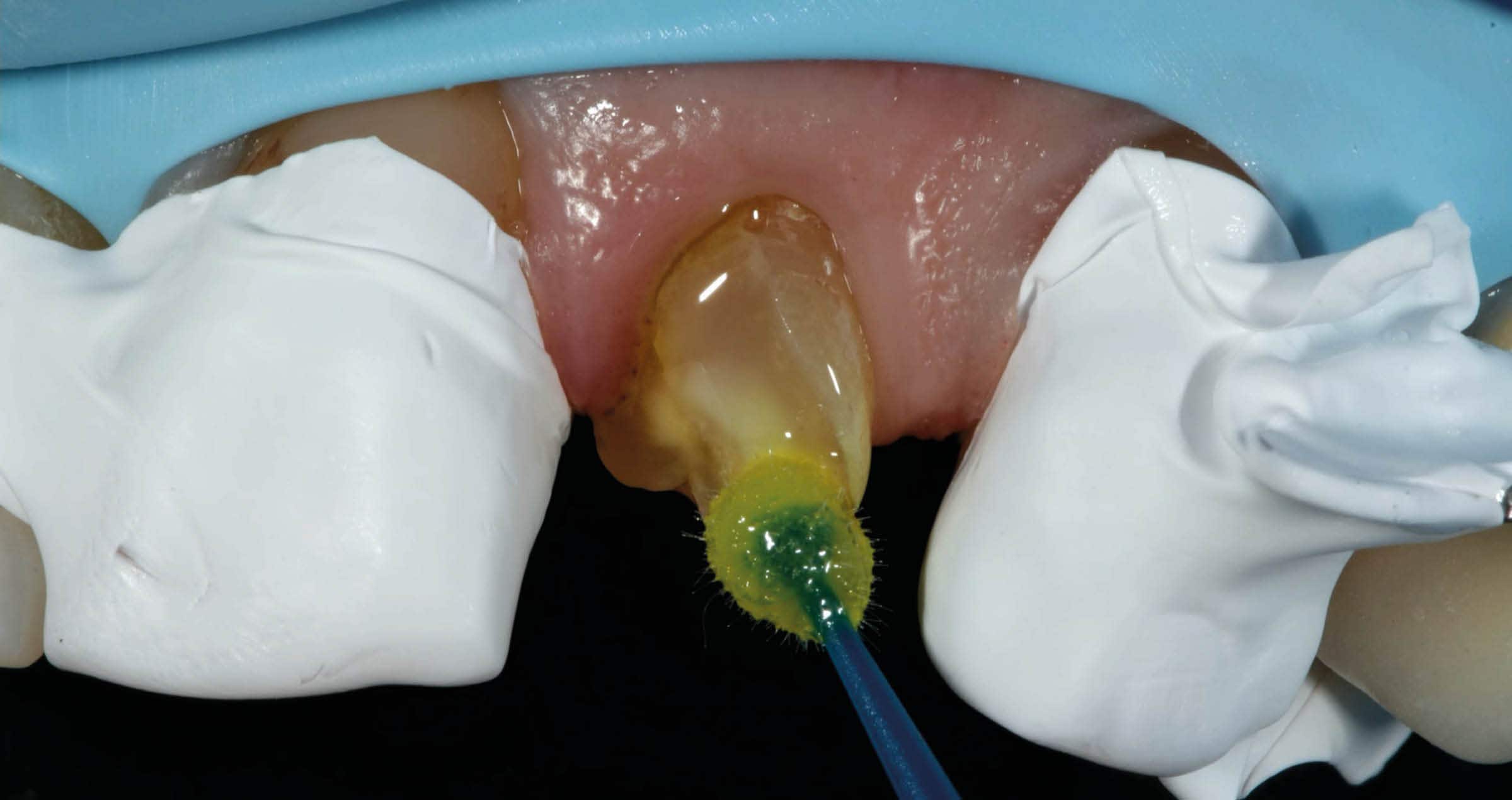
What followed was selective etching of the enamel on the tooth and the fragment for 15 seconds. Whenever selective enamel etching is the aim, it is essential to select an etchant with a stable (non runny) consistency – a property that is offered by K-ETCHANT Syringe (Kuraray Noritake Dental Inc.). Both surfaces were thoroughly rinsed and lightly dried before applying CLEARFIL™ Universal Bond Quick with a rubbing motion. This adhesive is really quick: Study results show that the bond established immediately after application is as strong and durable as after extensive rubbing into the tooth structure for 20 seconds.2,3 The adhesive layer was carefully air-dried to a very thin layer and finally polymerized on the tooth and on the fragment.


Fig. 7. Selective etching of the enamel of the tooth …


Fig. 8. … and the fragment with phosphoric acid etchant.


Fig. 9. Application …


Fig. 10. … of the universal bonding agent.


Fig. 11. Polymerization of the ultra-thin adhesive layer on the tooth …


Fig. 12. … and the fragment.


What happens to dentin in the selective enamel etch (or self-etch) mode?


After surface preparation or roughening, there is a smear layer on the dentin surface that occludes the dentinal tubules, forms smear plugs that protect the pulp and prevents liquor from affecting the bond. When self-etching the dentin with a universal adhesive, this smear layer is infiltrated and partially dissolved by the mild self-etch formulation (pH > 2) of the universal adhesive. At the same time, the adhesive infiltrates and demineralizes the peritubular dentin. The acid attacks the hydroxyapatite at the collagen fibrils, dissolves calcium and phosphate and hence enlarges the surface. Then, the 10-MDP contained in the formulation reacts with the positively loaded calcium (and phosphate) ions. This ionic interaction is responsible for linking the dentin with the methacrylate and thus for the formation of the hybrid layer.4,5


In the total-etch mode, the phosphoric acid is responsible for dissolving the smear layer and demineralising the hydroxyapatite. This leads to a collapsing of the collagen fibrils, which need to be rehydrated by the universal adhesive that is applied in the next step. Whenever the acid penetrates deeper into the structures than the adhesive, the collagen fibrils will remain collapsed. This will most likely result in clinical issues including post-operative sensitivity6.


When applying the adhesive system, a dental practitioner rarely thinks about what is happening at the interface7. However, every user of a universal adhesive should be aware of the fact that a lot is happening there. This is why it is so important to use a high-performance material with well-balanced properties and strictly adhere to the recommended protocols.


Fig. 13. Schematic representation of dentin after tooth preparation: The smear layer on top with its smear plugs occluding the dentinal tubules protects the pulp and prevents liquor from being released into the cavity.


Fig. 14. Schematic representation of dentin after the application of a universal adhesive containing 10-MDP: The mild self-etch formulation partially dissolves and infiltrates the smear layer, while at the same time demineralizing and infiltrating the peritubular dentin5.


In the present case, the tooth and the fragment now needed to be reconnected. For this purpose, CLEARFIL MAJESTY™ ES-Flow (A2 Low) was applied to the tooth structure. The fragment was then repositioned with a silicone index, held in the right position with a plier and light cured. To obtain a smooth margin and glossy surface, the restoration was merely polished. The patient presented after 1.5 years for a recall and the restoration was still in a perfect condition.


Fig. 15. Reconnecting the fragment with the tooth structure.


Fig. 16. Treatment outcome.


Why is it important to adhere to the product-specific protocols?


Universal adhesives contain lots of different technologies in a single bottle. While this fact indeed allows users to rationalize their clinical procedures, it also requires some special attention. As every highly developed material, universal adhesives need to be used according to the protocols recommended by the manufacturer. In general, materials may only be expected to work well on absolutely clean surfaces, while contamination with blood and saliva is likely to decrease the bond strength significantly. Depending on the type of universal adhesive, active application is similarly important, as is proper air-drying and polymerization of the adhesive layer. In addition, care must be taken to use the material in its original state, which means that it needs to be applied directly from the bottle to avoid premature solvent evaporation or chemical reactions. When adhering to these rules, universal adhesives offer several benefits from streamlined procedures to simplified order management and increased sustainability, as fewer bottles are needed and likely to expire before use.


Dentist:

DR. JOSÉ IGNACIO ZORZIN


Dr. José Ignacio Zorzin graduated as dentist at the Friedrich-Alexander University of Erlangen-Nürnberg, Germany, in 2009. He obtained his Doctorate (Dr. med. dent.) in 2011 and 2019 his Habilitation and venia legendi in conservative dentistry, periodontology and pediatric dentistry (“Materials and Techniques in Modern Restorative Dentistry”). Dr. Zorzin works since 2009 at the Dental Clinic 1 for Operative Dentistry and Periodontology, University Hospital Erlangen. He lectures at the Friedrich-Alexander University of Erlangen-Nürnberg in the field of operative dentistry where he leads clinical and pre-clinical courses. His main fields of research are self-adhesive resin luting composites, dentin adhesives, resin composites and ceramics, publishing in international peer-reviewed journals.

References


1. Van Meerbeek, B.; Yoshihara, K.; Van Landuyt, K.; Yoshida, Y.; Peumans, M. From Buonocore‘s Pioneering Acid-Etch Technique to Self-Adhering Restoratives. A Status Perspective of Rapidly Advancing Dental Adhesive Technology. J Adhes Dent 2020, 22, 7-34.
2. Kuno Y, Hosaka K, Nakajima M, Ikeda M, Klein Junior CA, Foxton RM, Tagami J. Incorporation of a hydrophilic amide monomer into a one-step self-etch adhesive to increase dentin bond strength: Effect of application time. Dent Mater J. 2019 Dec 1;38(6):892-899.
3. Nagura Y, Tsujimoto A, Fischer NG, Baruth AG, Barkmeier WW, Takamizawa T, Latta MA, Miyazaki M. Effect of Reduced Universal Adhesive Application Time on Enamel Bond Fatigue and Surface Morphology. Oper Dent. 2019 Jan/Feb;44(1):42-53.
4. Fehrenbach, J., C.P. Isolan, and E.A. Münchow, Is the presence of 10-MDP associated to higher bonding performance for self-etching adhesive systems? A meta-analysis of in vitro studies. Dental Materials, 2021. 37(10): 1463-1485.
5. Van Meerbeek, B., et al., State of the art of self-etch adhesives. Dental Materials, 2011. 27(1): 17-28.
6. Pashley, D.H., et al., State of the art etchand-rinse adhesives. Dent Mater, 2011. 27(1): 1-16.
7. Vermelho, P.M., et al., Adhesion of multimode adhesives to enamel and dentin after one year of water storage. Clinical Oral Investigations, 21(5): 1707-1715.


Cementering av indirekta protetiska ersättningar

Vissa företag använder i huvudsak basteknologier som utvecklats av andra för att förbättra sina produkter och för att introducera nya. Andra företag bedriver egen grundforskning och teknologisk utveckling. Är den skillnaden väsentlig för den som dagligen använder slutprodukten i sin praktik eller i sin laboratorieverksamhet? Svaret är ja - som företag med djup förståelse för underliggande komponenter, kemi och teknologier är vi kapabla att lösa existerande utmaningar och att snabbt och flexibelt svara upp till marknadens behov. Den här artikeln beskriver betydelsen av flera av de basteknologier som utvecklats av Kuraray Noritake Dental Inc. för cementering av indirekta protetiska ersättningar.


Adhesiv cementering då och nu


Möjligheten att fräsa fram dentala ersättningar ur flera olika sorters keramer har öppnat upp nya möjligheter för protetiken: högestetiska ersättningar kan framställas och installeras. Det är lätt att glömma bort eller undervärdera den roll som adhesiva cementeringssystem spelar i sammanhanget; den adhesiva cementeringen påverkar inte bara estetiken hos den translucenta tandfärgade protetiken, den banar också väg för en mindre invasiv preparationsteknik och för smäckrare utformningen av protetiken. De cementeringssystem som först fanns på marknaden och som möjliggjorde en kemisk bindning mellan tand och protetik var olyckligtvis väldigt teknikkänsliga och erbjöd ett mindre pålitligt resultat ur perspektiven ”hållbarhet” och ”långvarighet” samtidigt som arbetsprocessen var väldigt komplicerad. Den teknologi som utvecklats av Kuraray Noritake Dental Inc. bidrog signifikant till att förbättra cementeringens prestanda över tid och förenklade handhavandet avsevärt.


Optimering av bondingens långtidsprestanda


För att uppnå bondingresultat som varar över tid; speciellt bonding till dentin i de tidiga cementeringssystemen, beslutade Kuraray - ett moderföretag till Kuraray Noritake Dental Inc. - på 1970-talet att fokusera på utvecklingen av en mer kraftfull monomer. Som ett första steg på vägen mot fulländning, introducerades 1976 fosfatmonomeran Phenyl-P. Fem år senare hade fortsatta ansträngningar att förbättra och förfina molekylstrukturens hos Phenyl-P lett fram till introduktionen av den populära MDP-monomeren, som har förmågan att etablera en väldigt stark och hållbar bindning till emalj, dentin, metall och zirkonia.


Det faktum att MDP-monomeren fortfarande utgör en komponent i alla de adhesiver och adhesiva cementeringssystem som tillverkas av Kuraray Noritake Dental Inc. - och som också används av andra tillverkare för att optimera bindningsstyrka och hållbarhet för deras produkter - understryker uppfinningens sinnrikhet. I jämförelse med MDP som framställs av andra tillverkare står originalet - Original MDP Monomer från Kuraray Noritake Dental Inc. - ut på grund av sin ojämförbara renhetsgrad. Oberoende studier visar att graden av renhet har en positiv effekt på bindningen1. Genom att sin stabilitet i fuktiga miljöer, bidrar MDP-monomeren till en högre grad av stabilitet och pålitlighet hos de produkter som innehåller den.


Olika MDP-monomerer skiljer sig åt i renhetsgrad och bindningsförmåga.

Tre experimentella självetsande primers preparerades med 15 wt%. 10-MDP tillhandahölls av olika tillverkare: KN (Kuraray Noritake Dental), PCM (Germany) eller DMI (Designer molecules Inc., USA). Data med tillstånd av Dr. Kumiko Yoshihara.


För att adhesiva resincementeringssystem ska kunna ge en stark bindning med utomordentlig kantförsegling räcker det dock inte med att materialet innehåller en adhesiv monomer. Effektiv polymerisation är också en nödvändighet - även om en sådan inte alltid är så enkel att uppnå. För att bidra till en effektiv ljushärdning - såväl som härdning utan ljus - av PANAVIA™ V5, har Kuraray Noritake Dental Inc. utvecklat Touch-Cure Technology. Nyckeln till denna teknologi är en nyligen utvecklad högaktiv accelerator för polymerisation i PANAVIA™ V5 Tooth Primer. Acceleratorn har förmågan att inte brytas ned av MDP-syran. Så fort PANAVIA™ V5 Paste appliceras på tandytan som behandlats med primer främjar monomeren polymerisation med start från gränssnittet mellan tanden och cementet. I PANAVIA™ Veneer LC – ett ljushärdande resincementsystem som fungerar med samma primer – uppvisar acceleratorn i PANAVIA™ V5 Tooth Primer samma verkningsmekanismer. Den bidrartill polymerisationen av det adhesiva gränssnittet, medan PANAVIA™ Veneer LC Paste erbjuder förnämlig ljusstabilitet och är ljushärdande.


Detta fenomen utvärderades till exempel i PANAVIA™ F2.0 - föregångaren till PANAVIA™ V5. Resultatet av studien: PANAVIA™ F2.0 uppvisade mycket bättre egenskaper avseende kantförsegling än andra utvärderade cement2. Denna dokumenterat säkra försegling av gränsskiktet leder till lägre risk för marginalt läckage, till en mycket hög polymerisationsgrad också i fall där självhärdning (härdning utan ljus eller där ljuset blockeras av det restorativa materialet) använts och ger därför en synnerligen stark bindning.


Ytterligare en fördel som kommer sig av införlivandet av polymerisationsacceleratorn är dess funktion som ett starkt reduktionsmedel. Den neutraliserar natriumhypoklorit, som är vanligt förekommande som sköljlösning under endodontisk behandling, vilket innebär att dess negativa inverkan på bindningsstyrkan hos det därpå applicerade cementet elimineras.


Förenklad cementering av glaskeramer


Färre flaskor, färre steg och strömlinjeformade cementeringsmetoder: det var syftet med det tidiga 2000-talets utvecklig och marknadsföring av självadhesiva resincement. De flesta av dessa produkter har dock ett begränsat indikationsområde. De fungerar bra tillsammans med zirkonia, metall, emalj och dentin, men antingen rekommenderas de inte, eller kräver extra silanisering för att kunna användas tillsammans med glaskeramer. PANAVIA™ SA Cement Universal som innehåller MDP är annorlunda på grund av en annan märkesskyddad teknologi från Kuraray Noritake Dental Inc.: LCSi- Monomer, ett silankopplingsmedel bestående långa kolkedjor. Den här monomeren bildar en stark kemisk bindning med resinkomposit, porslin och silikatkeramer (som litiumdisilikat); därmed elimineras behovet av ett separat silan (primer eller adhesiv). Genom att utnyttja fördelarna med denna teknologi skiljer PANAVIA™ SA Cement Universal helt klart ut sig från andra självadhesiva resincement som ett äkta singelkomponentscement - till och med för glaskeramiska ersättningar.


Om så önskas kan produktens bindningsstyrka ökas genom användning av den populära universaladhesiven CLEARFIL™ Universal Bond Quick med Rapid Bond Technology. Denna teknologi har utvecklats av Kuraray Noritake Dental Inc. för att lösa problem relaterade till den långsamma vätningen av tandsubstans - särskilt vått dentin - som är typisk för universaladhesiver. För att kunna uppnå ordentlig vätning behöver dessa adhesiver aktivt gnuggas in i tandstrukturen under en relativt lång tid, alternativt måste användaren vänta en god stund innan skiktet kan ljushärdas. Det märkesskyddade Rapid Bond Technology, som består av Original MDP-monomer i kombination med hydrofila amidmonomerer, bidrar till en hög affinitet till vatten och ger en snabb och djup vätning av vått dentin. Följden blir en kortare applikationstid och enklare handhavande - utan negativ påverkan på bindingens kvalitet.


Slutsats


Teknologier utvecklade av Kuraray Noritake Dental Inc. har bidragit stort till förbättrade bondingegenskaper hos adhesiva cementeringssystem och en universell användning av självadhesiva resincemet. Företaget erbjuder därför ettströmlinjeformat sortiment av högpresterande resincement för alla användare - i alla kliniska situationer. För enklare procedurer med färre misstag och för hållbara estetiska ersättningar som varar över tid krävs cementeringssystemsystem med färre komponenter och färre arbetsmoment. Förutom de teknologiska fördelarna erbjuder dessa produkter många andra fördelar: detaljerade beskrivningar finns online på kuraraynoritake.eu.   


Referenser


*) Functional monomer impurity affects adhesive performance.; Yoshihara K, Nagaoka N, Okihara T, Kuroboshi M, Hayakawa S, Maruo Y, Nishigawa G, De Munck J,
Yoshida Y, Van Meerbeek B. Dent Mater. 2015 Dec;31(12):1493-501.
**) Touch-Cure Polymerization at the Composite Cement-Dentin Interface.; Yoshihara K, Nagaoka N, Benino Y, Nakamura A, Hara T, Maruo Y, Yoshida Y, Van Meerbeek
B.J Dent Res. 2021 Aug;100(9):935-94


Förenklade arbetssätt fördirekta restaurationer

Vissa företag använder i huvudsak basteknologier som utvecklats av andra för att förbättra sina produkter och för att introducera nya. Andra företag bedriver egen grundforskning och teknologisk utveckling. Är den skillnaden väsentlig för den som dagligen använder slutprodukten i sin praktik eller i sin laboratorieverksamhet? Svaret är ja! Producenter som utvecklar allting från grunden har vanligen en djupare förståelse av sina produkter och sina tillverkningsprocesser. Det gör det lättare för förtaget att modifiera specifika egenskaper, att lösa problem och att svara upp till marknadens behov. Den här artikeln beskriver betydelsen av flera av de basteknologier som utvecklats av Kuraray Noritake Dental Inc. för arbetsflödet vid direkt cementering av protetiska ersättningar


Direkta fyllningar – från komplext till enkelt

Ett av de vanligaste sätten att behandla kariesskadade tänder är direkta lagningar med dentala adhesiver med hög prestanda och med resinkompositer. Förutsatt att materialen är väl valda, kan man nuförtiden skapa ett vackert och hållbart resultat med enbart en flaska universaladhesiv och en eller två kompositer med olika opacitetsgrad. Så har dock inte alltid varit fallet. Länge var metoderna för att skapa direkta ersättningar ganska komplicerade; adhesiverna var teknikkänsliga system med många flaskor och flera tidskrävande behandlingssteg. Komposita fyllningsmaterial kunde bara fås att efterlikna naturliga tänder genom en exakt kombination av många olika färger och opacitetsgrader. Även om den komplicerade proceduren utfördes korrekt var ändå risken för mikroläckage, missfärgning och sekundärkaries relativt hög. Kuraray Noritake Dental Inc. fokuserade ganska tidigt på att lösa dessa problem; inledningsvis genom att använda Original MDP Monomer, som utvecklats 1981.


Optimering av adhesionsegenskaperna

Original MDP Monomer adresserade frågan rörande den begränsande långtidsprestandan hos de adhesiva systemens bindningsförmåga. Den hydrofila fosfatgruppen i MDP formerar en särskilt stark och långlivad kemisk bindning med kalcium som återfinns i hydroxylapatit, som är den huvudsakliga komponenten i emalj och dentin. Bindningen mellan MDP och kalcium utgör basen för ett stabilt, starkt och hållbart hybridlager. I kombination med resinet i bindemedlet resulterar det - efter ljushärdning - till en tät försegling av kaviteten. Fram till dagens datum är MDP fortsatt en absolut nödvändig komponent i varje produkt från Kuraray Noritake Dental Inc., och den är nyckelkomponenten som gjort CLEARFIL™ SE Bond till de självetsande adhesivsystemens gyllene standard.

Original MDP- monomeren skapar en stark kemisk bindning till emalj, dentin, metaller och zirkonia.

Övertygade om att dentala adhesiver skulle kunna utgöra mer än bra en stark och hållbar bindning började Kuraray Noritake Dental Inc. fokkusera på att lösa ett annat problem: risken för demineralisering och sekundärkaries som orsakas av resterande bakterier i kaviteten. Med utgångspunkt i sina erfarenheter av att utveckla andra adhesiva monomerer, uppfann Kuraray Noritake Dental Inc. MDPB-monomeren som har en antibakteriell och rengörande effekt i kaviteten. Till skillnad från antibakteriella medel, som kan försämra bindningsstyrkan hos en senare applicerad adhesiv, dödar MDPB-monomeren kvarvarande bakterier utan att påverka bindningsegenskaperna. Den ingår i den självetsande tvåkomponents-adhesiven i primersytemet CLEARFIL™ SE Protect och binds av polymerisation.


Den bakteriedödande egenskapen hos MDPB antas vara liknande den som kan iakttas hos CPC (Cetylpyridiniumklorid), som återfinns i många tandkrämer och produkter för munskölj.


Medan de självetsande tvåkomponentsadhesiverna redan har förenklat den adhesiva processen, utgör enkomponentsadhesiven av univeraltyp ”det lilla extra”. Det är en utmaning att föra samman ingredienser, som tidigare fördelats ut i ett flerkomponentssystem, i en och samma behållare, utan att riskera produktens stabilitet. Modern teknologi gör det nu möjligt. För att försegla ytan så fort som möjligt efter applicering måste momoneren snabbt och effektivt väta tandsubstansen. Vätningen brukar dock fördröjas av monomerer som behöver tid för att väta tandstrukturen – särskilt vått dentin – och ibland behöver de till och med gnuggas in. Därför har Kuraray Noritake Dental Inc. fokuserat på att utveckla Rapid Bond-teknologin. Den består av Original MDP-momomeren i kombination av nyligen utvecklade hydrofila korslänkade amidmonomerer och är integrerad i CLEARFIL™ Universal Bond Quick. De hydrofila amidmonomererna bidrar till en snabb, djup och fullständig vätning av dentinet och bildar vid härdning ett tätt, korslänkat polymernät som garanterar en stark och hållbar bindning. Både väntetid och arbetstid blir därigenom väsentligt förkortad och efter ljushärdning etableras en tättslutande och stabil försegling av kaviteten.

PREPARERAT DENTIN MED ”SMEAR LAYER”
På grund av dess hydrofila egenskaper behövs ett mycket hydrofilt bindemedel för att uppnå optimal vätning av dentinet.

DENTIN SOM BEHANDLATS MED CLEARFIL™ UNIVERSAL BOND QUICK.

Under härdning bildar CLEARFIL™ Universal Bond Quick ett höggradigt korslänkat nätverk av polymer. Tack vare nätverket är bindningens upptagning av vatten mycket liten och ger en stabil och bestående lagning.


Tack vare sina hydrofila egenskaper (vattenaffinitet) ger amidmonomererna i Rapid Bond Technology en mycket god vätning. Efter ljushärdning uppvisar bindningen låg upptagning av vatten och därför mycket hög motståndskraft mot åldrande.


Optimerar direkta fyllningar

Kombinerar flera lager, färger och opacitetsgrader: Användningen av mycket komplicerade tekniker för att genom olika lager skapa naturtrogna kompositfyllningar hör lyckligtvis – i de flesta kliniska situationer - till det förgångna. Anledningen: Högutvecklade resinkompositer som sömlöst smälter in bland den omgivande tandstrukturen. För att kunna tillhandahålla denna gynnsamma fördel har Kuraray Noritake Dental Inc. utvecklat sin varumärkesskyddade Light Diffusion Technology (LDT). Teknologin är inkorporerad i speciella förpolymeriserade fillerpartiklar vars funktion kan liknas vid tusentals mikroprismor som överför och bryter ljus och färg från den omgivande tandstrukturen. Fillerpartiklarnas optimala storlek, spridningen och ljusbrytningen i förhållande till matrisen ger en naturlig blandning av oöverträffat slag. Hela kompositserien CLEARFIL MAJESTY™ innehåller den varumärkesskyddade fillerteknologin. I det senaste tillskottet – CLEARFIL MAJESTY™ ES-2 Universal – använder Kuraray Noritake Dental Inc. nästa nivå – LDT - som möjliggör användning av en end färg med förenklad färgbestämning: den finns tillgänglig i två anteriora färger och i en färg för posteriort bruk, men smälter in så fint att den matchar praktiskt taget alla fäger i VITA classical A1-D4 shade guide.


Brytning och överföring av ljuset på rätt sätt ger fantastiska optiska egenskaper: ljusspridande partiklar i CLEARFIL MAJESTY™ ES-2 fån Kuraray Noritake Dental Inc.


Eftersom fantastiska synbara resultat inte bara är beroende av integration av de optiska egenskaperna och osynliga marginala gränser har Kuraray Noritake Dental Inc. också utvecklat optiska partiklar som sköter resten av jobbet – en naturlig ytglans och långvarig effekt av ytpolering. Lösningen som är integrerad i CLEARFIL MAJESTY™ ES Flow, med dess tre olika grader av flytbarhet, heter Submicron Filler Technology, och består av blanka partiklar i submikron storlek. Dessa partiklar är så små att ljusreflektionen uppvisar en stor naturtrogenhet– även efter slitage. Kuraray Noritake Dental Inc.’s enastående silanteknologi används både för att föra samman miljoner av de submikronsmå partiklarna och för att att hålla dem samman över tid. Det borgar för en hög fyllnadsgrad i de lågviskösa kompositerna och begränsar vattenupptagningen som annars skulle leda till försämring av den härdade kompositen. Den perfekta balansen mellan de små, blanka partiklarna, de ljusspridande partiklarna, resinmatrisen och den varumärkesskyddade silanteknologin ger en perfekt kombination av mekaniska och optiska egenskaper.

Exempel på ersättning framställd med en färg - CLEARFIL MAJESTY™ ES-2 Universal.


Sammanfattning

Adhesiva monomerer, fillerteknologier och silanteknologi ger en pålitlig kombination av partiklar, kluster och resinmatris: Kuraray Noritake Dental Inc. är definitivt en pålitlig expert på området adhesiva ersättningar. De varumärkesskyddade teknologierna som utvecklats under de senaste årtiondena har helt klart bidragit till bättre resultat för direkta ersättningar över tid, samt till att det blivit enklare att uppnå pålitliga och estetiska resultat.


Achieving maximum quality in a minimum amount of time

Interview with Andreas Chatzimpatzakis


Fewer bakes, fewer ceramic powders - there is clearly a trend toward simplification in the production of zirconia-based prosthetic work. This is also true for implant-based restorations, which often involve gum parts. DT Andreas Chatzimpatzakis, international trainer for Kuraray Noritake Dental Inc., and the owner of ACH Dental Laboratory in Athens, Greece, shares his approach to high aesthetics in implant prosthodontics in the following conversation.


You are a user of the CERABIEN™ ZR portfolio from the outset. When did you test the products for the first time and why?


Well, it was many years ago when I finished my very first zirconia-based restoration. The reason to test CERABIEN™ ZR was that when I asked the dental technician who had milled the framework which porcelain system to use. He suggested to use CERABIEN™ ZR, which I did. I was immediately impressed by the system and by the outcome I was able to achieve on the first attempt.


Did you ever test any other porcelain systems for ceramic layering?


Yes. Before I becoming an international trainer for Kuraray Noritake Dental Inc., I had the opportunity to test many other porcelain systems for layering on zirconia. Based on this experience, I can say that CERABIEN™ ZR is unique and the best system I have ever used. The reason is that its translucency and chroma are extremely close to natural teeth. In addition, due to a controlled firing shrinkage, a One-Bake Technique may be employed even in long-span restorations.


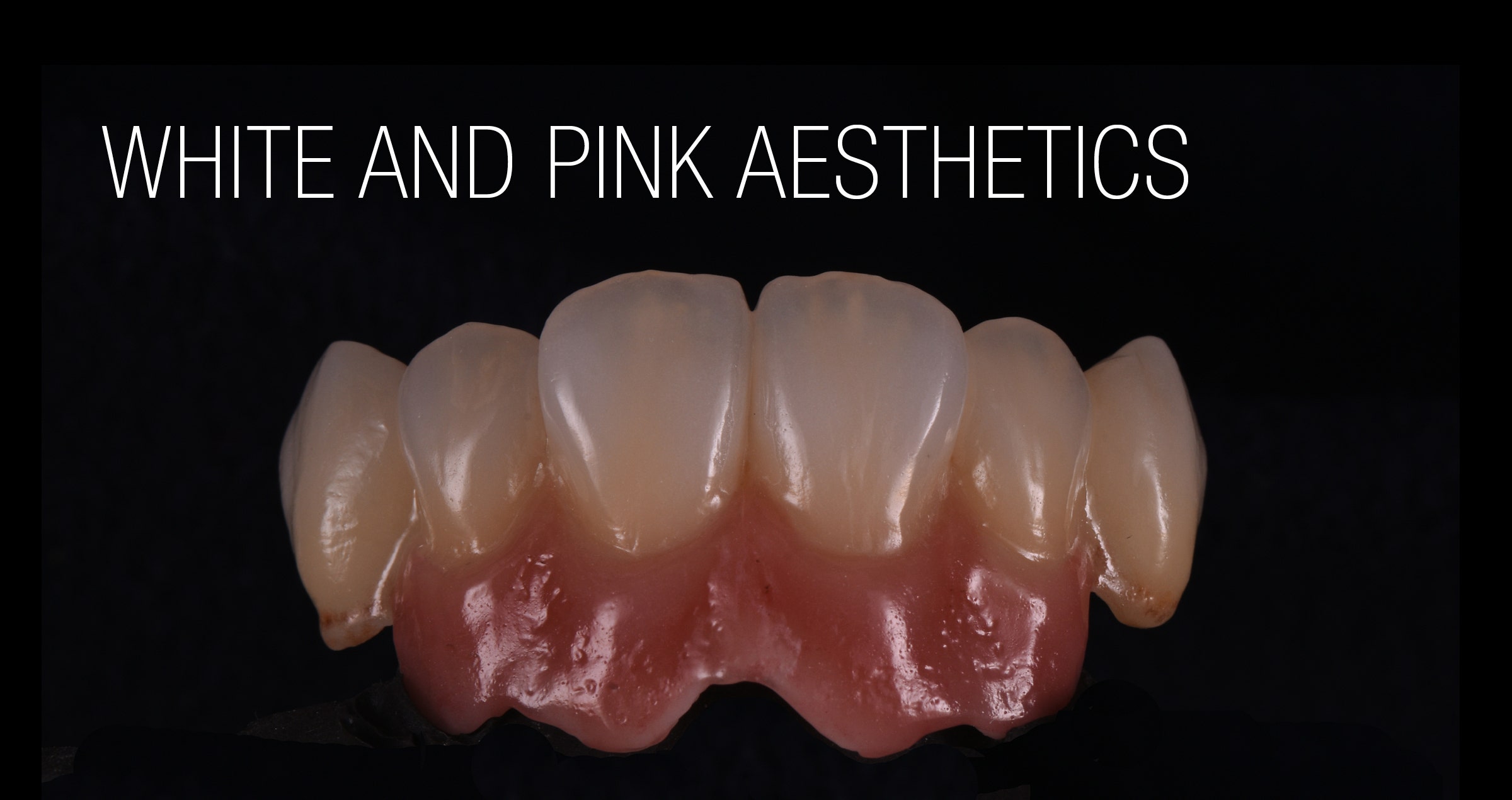
Your hands-on demonstration at the Kuraray Noritake Dental booth during the IDS 2023 in Cologne focused on White and Pink Aesthetics achieved with CERABIEN™ ZR. Is there a specific concept you use?


Nowadays, everyone producing dental restorations – no matter whether based on natural teeth or on implants – is confronted with increasing aesthetic demands of patients and dental practitioners. The high demands are developed because life-like restorations and cosmetic dental treatment outcomes are presented everywhere in the web and on social media. The showcased quality of outcomes is simply expected, even if the financial budget is limited. To be able to fulfil these demands in the field of implant-based prosthodontics, I have developed an approach that allows me to achieve high aesthetics with little effort. My concept is based on using not too many ceramic powders. For extra chroma and special characterization, I rely on the power of the internal live stain technique, first introduced by Hitoshi Aoshima-sensei.


Please summarize the most important details of your presentation.


The first important detail is the design and characterization of the framework. Before sintering, I apply Esthetic Colorant for KATANA™ Zirconia. After the sintering process, shade base stain and internal stains are mixed and applied. In this way, I create a nice canvas that helps me to achieve a life-like result with only a few selected ceramic powders. In most cases, three to five powders are enough to produce a great result. Among the powders used most frequently are Opacious Body, Body, LTX, Mamelon and CCV. After the first bake and a little grinding, I make use of internal stains again. They offer support in the controlling of the chroma and the integration of special characteristics. When this step is completed, the final build-up is done with one or two ceramic powders, most of the times LT1 and Enamel or LT0. Pink aesthetics are usually created with Tissue 1, 3 and 5. For the free gingiva, LT Coral is my go-to solution. The major goal is always to obtain maximum quality in a minimum of time. To achieve this, a good knowledge of the materials and of course practicing – on both, porcelain build-up and morphology – are strictly required.


Fig. 1. Complex implant-based restoration: Framework design.


Fig. 2. Esthetic Colorant …


Fig. 3.  … applied prior to the final sintering procedure.


Fig. 4. Appearance after sintering.


Fig. 5. Final outcome.


Are there any concrete tips and tricks you would like to share?


For the characterization of the framework, I mix the internal stains with shade base stain powders; mostly with SS Fluoro. For the first bake, especially when the restoration is large and the amount of ceramic to be applied huge, I reduce the heating rate up to 38 degrees per minute. I also increase the drying process up to 17 or even 20 minutes depending to the restoration. Experience shows that these measures optimize the aesthetic outcomes.


You often mention that it is extraordinarily important to understand the morphology of natural teeth to be able to produce beautiful restorations. Why is this the case?


A successful prosthetic restoration needs to offer proper function and aesthetics. Function means a precise fit, perfect contact points and occlusion, a proper emergence profile and interproximal embrasures for self-cleaning etc. All this is described by the term morphology. Aesthetics, on the other hand, is guided by shape and colour. The effort required to establish a proper morphology is much higher (about 70 percent of the total work) than the effort involved in obtaining the right translucency, opalescence and chroma.


What instruments do you use to imitate the morphology of natural teeth and how do you do it?


I usually make use of the Optimum™ Spring Ceramic Brush Size 8 (MPF Brush Co.), stones and diamond burs for detailed grinding and carving after the final bake. I studied morphology at the Osaka Ceramic Training Centre in Japan with Shigeo Kataoka-sensei. According to him, a key factor in creating a perfect macro and micro morphology lies in the shadows. To be able to take into account the interference of light and shadow during grinding, a light source is placed on one side of the restoration.


How many bakes do you need to produce highly aesthetic restorations?


It depends on the case, although in many situations, I nowadays opt for some kind of micro-layering. Lately, I have used micro-layering a lot with internal stain directly on the zirconia framework. In other cases, I do a quick first bake, then the internal staining, a final bake and glazing. For small or single-unit restorations in the posterior region, a One-Bake Technique is often sufficient. Even a Zero-Bake approach using Esthetic Colorant on a monolithic zirconia restoration may be appropriate here, and it is very convenient. If there is a restoration with high aesthetic demands – these are typically single anterior restorations – the technique I select depends on the shade. In some cases, using only the internal stain technique is enough to reach a high aesthetic level, while in other cases, additional steps need to be taken. To my mind, there is no single technique that fits all cases. As mentioned before, I try to achieve high aesthetics in a minimum of time.


Fig. 6. Clinical example of achieving high aesthetics in a minimum of time: Before …


Fig. 7. … and after crown placement.


Fig. 8. High aesthetics …


Fig. 9. … achieved in a minimum of time.


Did your approach change due to the availability of high-translucency zirconia materials with colour (and flexural strength) gradation?


Well, yes! My overall approach changed more to micro-layering. Several years ago, we needed to consider how to mask the framework and how to achieve translucency in areas with limited space. The problem was solved for single-unit and small anterior bridge restorations with the availability of KATANA™ Zirconia UTML and STML. With the introduction of KATANA™ Zirconia YML, a high-translucency material became available for long-span or implant-based restorations as well. We have strength and translucency all in one disc. In most of my cases, the framework material replaces the dentin with regard to morphology and shade. Hence, I need to focus on adding the enamel by applying the transparent and translucent powders. The powders of the internal stain technique are used to characterize the framework, and with a micro-layer of porcelain, the goal of creating an aesthetic restoration in the minimum of time is achieved. This is exactly why I am sure that micro-layering is the future.


What drives you to share your knowledge with others?


My passion! I love my work! And I love to see technicians become better and better. Dental technology is an exciting journey, a journey that begins when the first impression arrives in the dental laboratory, and it ends when the final restoration is cemented into the patient’s mouth. And this journey is so exciting because we change lives. We change people’s personalities, we give them back their smile, we give them back their self-respect. Consider that every day, every single moment working on our bench trying to imitate nature… there is nothing more exciting than that!!!


My approach as an instructor is to lead dental technicians to master the art of observing natural teeth. This is the way every individual will understand morphology and shade. You need no special talent to be a very good dental technician. You need to observe! Your eyes see, your mind understands, and your hands will follow.

Interview with Alexander Aronin

Alexander (Alek) Aronin is a master dental technician who dedicated himself to the creation of high-end, handmade porcelain restorations. He runs a dental laboratory and morphology school in Spain, and travels the world as a lecturer and teacher.


The greatest source of inspiration in his professional life is the book Collection of Ceramic Works by Hitoshi Aoshima. Through reading it in 1996, he discovered superb outcomes of working with dental ceramics. Moreover, the book’s content made him realize that the creation of handmade porcelain work is a result of special education and manual skills. Those who want to become masters need a lot of commitment and a striving for continuous improvement, which Alek shows in his work and his classes. We talked to him about his philosophy and his enthusiasm for the creation of lifelike restorations. He shared with us his perspective on the future of dental technology and gave some practical tips on how to achieve a high level of professional satisfaction.


Photo courtesy of Dennis Debiase


Alek, many dental technicians decide to focus on CAD/CAM technology and automated processing of dental ceramics. Due to improvements on the material side, a handmade porcelain layer is no longer necessary in many clinical cases. In this context, the manual refinement process is reduced to a minimum. Why did you decide to take a completely different path and focus on fully manual dental craftsmanship?


The shift towards automated processing is not a matter of choice for dental technicians rather, it is a natural response to the evolution of technology. The high-end manual refinement process remains unchanged. The human element, from communication to hand crafting among skilled individuals and demanding clients, has been a constant so far throughout history. This traditional connection remains stable for centuries.


Digitalisation is not the revolution in the dental industry, and I do not see the benefit of it in our narrow specialization yet. In the area we are working, we do all steps of our case faster, incomparably more precise and more profitable. But we are keeping eye on machines and waiting for a suitable one.


Machines and automated processes widely serve mass production businesses focused on fast, affordable and uniform results in a highly competitive field. Our goal and workflow are different - we provide individual work and personal attention to each of our partners and patients.


So, we do not compete with the production labs and do not interrupt each other, we coexist in parallel worlds as always. A small number of dentists and their patients will always demand personal attention and valued restorations and service of the highest quality.


Many dental technicians admire your work. Yet, you continue to strive for improvement. Why is this the case?


On one side, we are limited by static ceramic material used to mimic dynamic natural teeth that keep changing for a lifetime. On the other side, we are limited by our manual skills. I am still far away from my teachers and Japanese colleagues. My target is to improve the fabrication process. My goal is to achieve the simplicity and imperfectness like Aoshima-sensei.


Alek lecturing at the IDS 2023 in Cologne.
Photo courtesy of Dennis Debiase


We are enjoying the outcome, but prefer to focusing on the improvement of the process, and move on to create a better one. This is what I am learning in Japan, and this is what I teach my students.


Talking about learning: What are the most important aspects a dental technician who wants to improve his skills should have in mind when looking for a good teacher?


Manual skills are very important, but not the only aspect that should be taken into account. Every individual should be motivated and guided and this is a teacher’s job.


I love the traditional Japanese way of teaching and learning: The teachers are passionate, leading the way by evoking emotions and manual skills to bring the best out of every single student. My personal advice for dental technicians who want to become masters in the creation of lifelike high-end dental restorations is to select their teachers carefully and go to a private school or courses whenever they have the chance.


What are the most important tools a dental technician needs to use when trying to create high-end lifelike restorations?


I suggest to focus on four aspects:

  1. Documentary dental photography - required for documentation and communication with the dental office and patients using constant (once set and never changed) parameters of the photo equipment.
  2. Focus to the biomimetic additive dentistry. This is a minimum machine invasive field. Dental technicians and dentists should be able to develop a deep knowledge about clinical and lab-side procedures to be able communicate with each other.
  3. Mastering morphology and function (shape carving), and anatomy (internal staining), which comes with value control and mimicking fine tooth details for best integration in the mouth.
  4. Written communication (stop phone calls) is very important, this is the way how to exchange the information between the patient, clinic and lab by strict protocols.


I teach these complex skills in my morphology school and in many of my trainings worldwide. Focusing on the four aspects, a dental technician has a great chance to become a good specialist in a relatively short period of time in a narrow field.


Is material selection important for achieving great results?


I’ve been using Noritake ceramic for most of my life, and the reason is simple: Noritake created their EX-3 porcelain over 40 years ago, and it was so well-made that it has not needed any changes since.


This is showing their consistently high quality and creates unbroken succession in the valuable tradition of passing on techniques and knowledge.


Today, among different generations of dental technicians, we can use and share the same methods, vocabulary and abbreviations, powders, and temperature charts developed by our skilled teachers 30 to 40 years ago. This unique feature sets Noritake and Creation porcelains apart from all other brands and systems in the world.


The other Noritake porcelain I use quite frequently is CERABIEN™ ZR, which is also well-tried and tested and has even some more advantages than EX3.


Is there any final advice you would like to give?


To become a good professional, I suggest to developing in four parallel directions:

  1. Practicing on phantoms - fabricating cases and ceramic samples. It helps to experiment and practice with varied materials and techniques.
  2. Implementing the achieved techniques in clinical cases.
  3. Working with case presentation PowerPoint or Keynote: documenting the working steps in pictures and videos from beginning to end.
  4. Mastering the communication using e-mails. Constantly calibrate and adjust the information exchange process between the clinic and lab. Acquire deeper knowledge about the work of each other.


Good luck!


PANAVIA™: 40 years of success in adhesive luting

DID YOU EVER WONDER WHY THE PRODUCTS OF THE PANAVIA™ BRAND OFFER SUCH OUTSTANDING PERFORMANCE?


You probably know that they all contain the original MDP monomer developed in the early 1980s. It has attracted much attention because it is such an excellent adhesive monomer. This phosphate ester monomer forms a very strong bond to tooth structure, zirconia, and dental metals. It has been used in every PANAVIA™ product. In reality, however, other catalytic technologies and ingredients alongside MDP are important technological contributors supporting the performance of our cementation solutions.


INGREDIENTS AFFECTING THE POLYMERISATION REACTION


One of these decisive additional technologies and ingredients is the polymerization catalyst triggering the curing process. Different from the MDP monomer used in every PANAVIA™ product, the polymerization catalyst has been continuously improved since the introduction of PANAVIA™ EX in 1983. New versions have been developed for PANAVIA™ 21, PANAVIA™ Fluoro Cement and PANAVIA™ V5, for example. Another important component also affecting the curing process is the Touch Cure Technology used in two of the three major products of the current PANAVIA™ Portfolio: PANAVIA™ V5 and PANAVIA™ Veneer LC. This technology was first used in PANAVIA™ 21, which was launched in 1993. The contact of the chemical polymerization activator contained in the self-etching primer with the resin cement paste accelerates the polymerization of the cement from the adhesive interface, thus providing better adhesion of the resin cement. In developing PANAVIA™ V5, we reviewed the chemical composition of the existing PANAVIA™ products and updated it substantially. The Touch Cure technology has also been adopted for use in the case of PANAVIA™ V5 Tooth Primer and the concomitantly used PANAVIA™ V5 Paste. When cementing veneers with PANAVIA™ Veneer LC, we also use PANAVIA™ V5 Tooth Primer for conditioning teeth. This also involves the application of Touch Cure technology for achieving an adhesive connection with the tooth without compromising the working time of the cement paste.


ADDITIONAL ADHESIVE MONOMERS


Even in the field of adhesive monomers, we did not stand still: We developed the LCSi monomer, a long carbon chain silane-coupling agent which made possible to integrate the function of a ceramic primer in our universal self-adhesive resin cement PANAVIA™ SA Cement Universal. With its high level of hydrophobicity, this monomer provides stable, long-term bond strength. Generally speaking, it may be said that the reason bond durability may drop is a hydrolytic reaction damaging the chemical bond between the silica contained in the glass ceramics and the silane-coupling agent.


THREE PRODUCTS COVERING VIRTUALLY EVERY NEED


By combining these technologies and ingredients smartly, we have succeeded in developing a resin cement portfolio that covers virtually every need. With PANAVIA™ V5, PANAVIA™ SA Cement Universal and PANAVIA™ Veneer LC, it is possible to treat a wide variety of cases. The products allow for the luting of various types of restorations, prosthetic appliances and for the placement of posts and produce great outcomes if used properly according to the instructions for use.


PANAVIA™ V5 is the resin cement that has the widest range of uses among the three cement systems just mentioned. It covers almost all the intended uses of the other two cements. Therefore, it is possible for the dental practitioner to choose the right cement system for treatment, according to the cases arriving at the clinic and patient needs, from among these three major resin cement products: PANAVIA™ V5, a resin cement with a wide range of applications; PANAVIA™ SA Cement Universal, a simple and easy-handling self-adhesive resin cement; and PANAVIA™ Veneer LC when there is a need to bond laminate veneers.

EXPLORING NEW OPPORTUNITIES


The good thing about developing technologies in a company like Kuraray Noritake Dental is that their application is not limited to a certain product or product group. The R&D Department always carries out research on how to leverage the benefits of the technologies in other applications. Take, for example, KATANA™ Cleaner, which was released in 2019. This cleaning agent can be used to remove saliva, blood, temporary cement, or other contaminants that can adhere to the surfaces of teeth or prosthetic devices during trial fitting and temporary cementation of a prosthetic device. This unique product has been developed by taking advantage of the surfactant function of the MDP monomer.


CONCLUSION


Hence, it is mainly our long-standing knowledge and experience in the development of dental resin cements and adhesive solutions that provides for the excellence of the current PANAVIA™ Portfolio. We know how to improve on existing technologies, to never stop developing new ones and to continuously look for the best way to combine proven and new components to obtain the best possible outcomes. In the steps of the product development procedure, clinical tests are conducted and feedback from dental practitioners is gathered in order to take into account the extreme conditions found in the oral environment. In the past 40 years, this strategy has proven successful, and we are sure it will help us to develop many other innovative products that offer ideal support in striving to improve the oral health of patients.


Time to celebrate PANAVIA’s 40th anniversary

Interview with Mitsunobu Kawashima
Manager, Technology Division of Kuraray Noritake Dental Inc.


In 1983 – exactly 40 years ago – PANAVIA™ EX was introduced in Japan as the first product of the PANAVIA™ family and the first product containing the original MDP monomer. Since then, the PANAVIA™ family of resin cements has been continuously expanded by developing new resin-cement materials that are precisely adjusted to the contemporary demands of dental practitioners. The current line-up of easy-to-use, high-performance adhesive luting materials is globally available and used by dental practitioners with high quality standards.


We had a conversation with Mr. Mitsunobu Kawashima about PANAVIA™ EX as a ground-breaking innovation in the field of dental resin cements and subsequent steps toward the current well-balanced resin cement portfolio. He is currently responsible for the development of chair-side materials in the Technology Division of Kuraray Noritake Dental Inc. (Kuraray Noritake Dental) and has been part of the team developing the many products of the PANAVIA™ family for more than 30 years.


The resin cements developed by the company before the introduction of PANAVIA™ EX contained the adhesive monomer Phenyl-P. Why did you decide not to use Phenyl-P in the new formulation?


At the time we decided to develop PANAVIA™ EX, we were engaged in the development of new adhesive monomers to replace Phenyl-P. The main aim of this project was an improvement of our products’ bond strength to metal alloys and dentin. Among the new adhesive monomers being developed, we decided to use the MDP monomer, because it features excellent bonding to tooth structure and metal alloys used in dentistry, as well as superb resistance to water. Incidentally, our first products to contain the original MDP monomer were the resin cement PANAVIA™ EX and the bonding agent CLEARFIL™ NEW BOND.


Where did the name PANAVIA™ come from?


The name “PANAVIA™” is a compound word consisting of “PAN” and “VIA”. The former is of Greek origin, and the latter is of Latin origin. “PAN” means “everything” and “VIA” means “way” or “method”. Consequently, the name “PANAVIA” describes a “method for bonding everything”; it represents our desire to have products launched under the umbrella of the PANAVIA™ brand recognized as dental materials that can bond to all types of restoration and tooth structure.


Would you please tell us the story behind the development of PANAVIA™ EX?


Back in the early 1980s, it was indispensable for us to develop new adhesive monomers as a part of our project to develop a new resin cement with unprecedented adhesive properties. In this context, we conducted a comprehensive literature search for compounds thought to be involved in adhesion. At the same time, we carried out a variety of R&D activities, including basic research to quantitatively clarify the relationship between the molecular structures of monomers and their adhesive qualities, synthesis trials of various monomers and bond strength tests. Consequently, we succeeded in developing the MDP monomer that had the physical properties we were working to obtain. Following the development of that MDP adhesive monomer, we were finally able to create PANAVIA™ EX in our laboratory. In early 1982, we completed the first prototype of PANAVIA™ EX for external evaluation and asked a dental college in Japan to evaluate it. He found that the prototype cement might cure too quickly in clinical use. This was due to the fact that we had not taken into account the difference between room temperature and intraoral temperature, which has a huge impact on the curing time. This error led us to recognize how important clinical evaluations are during the development of dental materials. To this day, we continue to place great importance on the opinions of clinicians whenever a new material is being developed.


The name “PANAVIA™” is a compound word consisting of “PAN” and “VIA”. The former is of Greek origin, and the latter is of Latin origin. “PAN” means “everything” and “VIA” means “way” or “method”.


What were the key technological features of this new PANAVIA™ EX cementation system?


The system had five key features: Appropriate film thickness, appropriate flow properties of the paste, radiopacity, improved bond strength and improved surface-cure characteristics. At the time that PANAVIA™ EX was developed, a film thickness of 30 μm or less was desired for luting cements. The largest components we wanted to use in the formulation – the silica filler particles present in our composite resin – had a maximum particle diameter of 50 μm. We were able to achieve a film thickness of 30 μm or less by significantly extending the silica grinding time, which made the filler particles much finer. An appropriate paste consistency – a low level of viscosity and good flowability – was achieved by using low-viscosity monomers for PANAVIA™ EX. In order to make the cement radiopaque, we dispersed radiopaque filler within the powder component. In fact, we were convinced that it was essential to be able to check for the presence of excess cement under the gingival margins after a restoration was placed. The increase in bond strength was achieved by blending the MDP monomer into the liquid component.


What about the surface-cure characteristics of the cement?


Every dental practitioner knows that the surface of resin cements must be protected from oxygen in the air in order to cure properly. For this purpose, we developed OXYGUARD, a water-soluble gel material. It is applied to the restoration margins to cover the unpolymerized resin cement surface and protect it from exposure to oxygen. In this way, the formation of an oxygen inhibition layer – a layer of uncured resin on the surface that compromises the marginal integrity of the restoration – is prevented and an intact, fully polymerized cement surface can develop. In this way, the use of OXYGUARD has contributed greatly to improving the cure characteristics of cements.


What do today’s PANAVIA™ products and PANAVIA™ EX have in common?


The PANAVIA™ family of products, including PANAVIA™ EX, share the concept of “being a resin cement product that opens new dimensions in dentistry”. PANAVIA™ EX was our first resin cement, and it was conceived as a cement for a new era. It can bond to tooth structure and dental metals very well, thanks to the use of the MDP monomer. This important adhesive monomer is still used today in many of our products. Ever since the launch of PANAVIA™ EX, Kuraray Noritake Dental has continued to take on new challenges and has developed many new products in the growing PANAVIA™ family. These include PANAVIA™ 21, a cement in paste form that has a self-etching primer as an accessory; PANAVIA™ Fluoro Cement, a dual-cure cement paste that releases fluoride; and PANAVIA™ F2.0, which can be used with an LED curing unit. The current portfolio consists of PANAVIA™ V5, which features the substantially improved bonding performance that was achieved after a major review of the basic composition of the series, PANAVIA™ SA Cement Universal, which works as a standalone product without separate primers, and PANAVIA™ Veneer LC, PANAVIA™ family’s latest product. The latter is a light-curing resin cement with the specialized purpose of bonding laminate veneers.


In retrospect, what did the introduction of PANAVIA™ EX mean to Kuraray Noritake Dental?


In the development of new resin cements, it is important to quickly respond to the rapidly changing trends in the market. Whenever new prosthodontic treatment concepts appear – like adhesive bridges as a minimally invasive treatment option replacing a single tooth – or new restorative materials are introduced, such as different types of ceramics – we need to check if our resin cement systems are compatible with the tasks that come with these changes, as well as possibly developing new ones. Following the launch of PANAVIA™ EX, we received a wide range of feedback from experts working at dental clinics and laboratories. We leveraged this feedback in subsequent development projects, always striving to stick close to the clinical setting. After Kuraray Medical Inc. merged with Noritake Dental Supply Co., Limited, this ethos was embedded in the work ethic of all employees of Kuraray Noritake Dental.


Among the new adhesive monomers being developed, we decided to use the MDP monomer, because it features excellent bonding to tooth structure and metal alloys used in dentistry, as well as superb resistance to water.


Can you give us a brief history of the PANAVIA™ EX journey to the currently available PANAVIA™ family portfolio?


Since the launch of PANAVIA™ EX 40 years ago, six different PANAVIA™ products have been released. Each of them has received high acclaim for their unique features, both in the Japanese resin cement market and abroad. For 40 years, we have been improving PANAVIA™ in response to the demands of the times. We developed a product with increased adhesion when non-retentive preparation designs (adhesive bridges) and smaller bonding surfaces (due to less invasive preparations) became popular. And for users concerned about secondary caries, we developed a material with fluoride-releasing properties. At the same time, we focused strongly on making dental cementation easier by finding ways to make our resin cements bond well to various types of crown-restoration materials, including precious metals and ceramics. During these drives towards improvement, “achieving reliable general luting” has always been at the forefront of our goals. We believe that the successful evolution of the PANAVIA™ brand owes a great deal to our incessant efforts to obtain a high level of bond strength to dentin after chemical polymerisation”.


How did the R&D department evolve over the years?


Initially, our efforts in the development of dental materials had focused on bonding agents and restorative composite resins. PANAVIA™ EX was just one of our new development projects and only a few staff members were assigned to that product. After PANAVIA™ EX was launched in 1983 and it was accepted worldwide, our product line-up of resin cements was expanded considerably. Consequently, we have increased the number of development staff assigned to resin cements and set up a special development team responsible for the self-adhesive resin cement product line that includes PANAVIA™ SA Cement Universal. As a result, the size of the team assigned to resin cements has expanded substantially.


How did production change?


When PANAVIA™ EX was launched in 1983, the product was manufactured exclusively for the Japanese market in a relatively small facility. Today, in order to produce all products of the PANAVIA™ family for the global market, we have automated our production facilities and increased the production equipment and systems dedicated to producing the resin cement paste. To support our product quality, we have also set up a quality control system that assures we turn out safe and high-quality products, drawing on over 40 years of technical knowhow and experience in the production of resin cements.


For 40 years, we have been improving PANAVIA™ in response to the demands of the times.


When did external researchers start showing interest in PANAVIA™?


The development of PANAVIA™ EX was carried out with the active participation of researchers at a dental college in Japan, involving such activities as performing basic adhesion tests and experimenting with clinical applications using adhesive bridges. At that time, resin cements that provided a strong bond to tooth structure or metal alloys were not widely used. I believe that overseas researchers were interested in the development of PANAVIA™ EX at a relatively early stage, for this reason.


What current concept do you have in mind as you continue developing your adhesive cements?


We have two basic central concepts: “Achieving greater bond strength” and “Delivering easier handling characteristics”. I think that the shades of resin cements are also devised in each product in order to get the most of the characteristics of aesthetic restorative materials. At our company, we have embodied “achieving greater bond strength” in PANAVIA™ V5, and “delivering easier handling characteristics” in PANAVIA™ SA Cement Universal. We will continue to explore the development and introduction of various new technological applications, to bring even higher performance products to market.


What do you think is the strength of Kuraray Noritake Dental’s R&D team?


We conduct R&D activities continuously, focusing on the development of luting materials. In our product development department, the same person is often responsible for one product category over long period of time. For example, I have been engaged in the development of PANAVIA™ products for much of my time with the company. The result is that each person in the development department can be said to be an expert in a certain category of products. They leverage the technical knowledge acquired in the past for the development of new products, resulting in entirely new discoveries.


Do you have any ideas about what the future of PANAVIA™ will be?


We will continue to focus on the development of even simpler, easier-to-use resin cements suitable for a wide range of applications – products based on the concept of universality. This should allow users to focus more on the actual treatment than ever before, while of course delivering a strong and durable bond between the tooth structure and the restoration.


MITSUNOBU KAWASHIMA


Optimizing functional and esthetic parameters in veneer cementation

By Dr. Clarence Tam, HBSC, DDS, AAACD, FIADFE


The use of both porcelain veneers to improve and restore the shape, shade and visual position of anterior teeth is a common technique in esthetic dentistry. The biomimetic aim in the restoration of teeth is not only the cosmetic domain, but also functional considerations. It is critical to note that the intact enamel shell of the palatal and facial walls with respect to anterior teeth are responsible for its innate flexural resistance. When dental structure has been violated by endodontic access, caries and/or trauma, every effort must be made to preserve the residual structure and strive to restore or exceed the baseline performance levels of a virgin tooth.


BACKGROUND


A 55 year old ASA II female with a medical history significant only for controlled hypertension presented to the practice for teeth whitening. It was foreseen that dental bleaching would not have an effect on the shade of a pre-existing porcelain veneer on tooth 1.2, and that this would need to be retreated following the procedure especially if the shade value changes were significant. The patient started with a baseline shade of VITA* 1M1:2M1; 50:50 ratio in the upper anterior region and 1M1 in the lower anterior region. Following a nightguard bleaching protocol with 10% carbamide peroxide worn overnight for 3-4 weeks, the patient succeeded in achieving a VITA* 0M3 shade in both upper and lower arches. As a result, there was a significant value discrepancy between the veneered tooth 1.2 and the adjacent teeth, and also increased chroma noted on the contralateral tooth 2.2 due to a facially-involved Class III composite restoration. This latter tooth also did not match the contralateral tooth in dimension and thus the decision was made to treat both lateral incisors with bonded lithium disilicate laminate veneers. The canine adjacent (2.3) featured localized mild to moderate cusp tip attrition, but the patient did not want to address this until following the currently-discussed veneers were placed. The goal of smile design at this stage is to ultimately establish bilateral harmony with the view to place an additional indirect restoration restoring the facial volume and cusp tip deficiency of tooth 2.3 in the near future.


PROCEDURE


A digital smile design protocol was not required for the initial intention, which was individual treatment of the lateral incisors, as slight variation is permitted in this tooth type, being a personality and gender marker of the smile. Prior to anesthesia, the target shade was selected using retracted photos featuring both polarized and unpolarized selections. The photographs were prepared for digital shade calibration by taking reference views with an 18% neutral gray white balance card (Fig. 1).


Fig. 1. Reference photograph taken with a 18% neutral gray card.


The basic body shade was VITA* 0M2 with an ingot shade of BL2. The patient was anesthetized using 1.5 carpules of a 2% Lignocaine solution with 1:100,000 epinephrine before affixing a rubber dam in a split dam orientation. The veneer on tooth 1.2 was sectioned and removed from tooth 1.2 and a minimally-invasive veneer preparation completed on tooth 2.2 (Fig. 2). Partial replacement of the old composite resin restoration was completed on the mesioincisobuccopalatal aspect of tooth 12 with the intact segment maintained. Adhesion to old composite was achieved using both micro particle abrasion and a silane coupling agent (CLEARFIL™ CERAMIC PRIMER PLUS, Kuraray Noritake Dental Inc.). Margins were refined and retraction cords soaked in an aluminum chloride solution and packed. Preparation stump shades were recorded. Final impressions were taken using both light and heavy body polyvinylsiloxane in a metal tray. The patient was provisionalized and sent away with instructions to verify the shade at the laboratory at the bisque bake stage. The models prepared by the laboratory verify the minimally-invasive nature of the case.


Fig. 2. Veneer preparation tooth 1.2, 2.2.


On receipt of the case, the patient was anesthetized and the provisionals removed. The preparations were debrided and prepared for bonding by abrading the surfaces using a 27 micron aluminum oxide powder at 30-40 psi. The veneers were assessed using a clear glycerin try-in paste (PANAVIA™ V5 Try-in Paste Clear, Kuraray Noritake Dental Inc.). Retraction cords were packed and the intaglio surface of the restorations treated using a 5% hydrofluoric acid for 20 seconds prior to application of a 10-MDP-containing silane coupling agent (CLEARFIL™ CERAMIC PRIMER PLUS, Kuraray Noritake Dental Inc.) (Fig. 3). The tooth surface was etched using 33% orthophosphoric acid for 20 seconds and rinsed. A 10-MDP-containing primer was applied to the tooth (PANAVIA™ V5 Tooth Primer, Kuraray Noritake Dental Inc.) (Fig. 4) and air dried as per manufacturer’s instructions. Veneer cement was loaded (PANAVIA™ Veneer LC Paste Clear, Kuraray Noritake Dental Inc.) (Fig. 5) and the veneer seated. The excess cement featured a non-slumpy character and maintained the veneer well in place during all margin verification exercises prior to a 1 second tack cure (Fig. 6).


Fig. 3. CLEARFIL™ CERAMIC PRIMER PLUS applied to intaglio surfaces of veneers.


Fig. 4. PANAVIA™ V5 Tooth Primer application to etched tooth surfaces.


Fig. 5. PANAVIA™ Veneer LC Paste Clear shade loaded onto prepared intaglio surfaces of veneers.


Fig. 6. PANAVIA™ Veneer LC Paste immediately after seating. Note the viscous, non-slumpy nature of the cement, which allows for ease of removal under both wet and gel-phase options.


The cement was rendered into a gel state, which facilitated “clump” or en masse removal of cement with minimal cleanup required (Fig. 7). The margins were coated using a clear glycerin gel prior to final curing to eliminate the oxygen inhibition layer (Fig. 8).


Fig. 7. Excess cement removal after tack curing for 1 second.


Fig. 8. Final curing of veneers from both palatal and facial aspects simultaneously.


The margins were finished and polished to high shine and the occlusion of the restorations verified as conformative. The post-operative views show excellent esthetic marginal integration (Fig. 9).


Fig. 9. Post-operative esthetic integration of veneers on 1.2 and 2.2.


On polarized photograph reassessment, the restorations are well-integrated into the new smile esthetically and functionally (Fig. 10), now awaiting esthetic augmentation of tooth 2.3 to match the contralateral canine.


FINAL SITUATION


Fig. 10. Final result with polarized photography on reassessment.


RATIONALE FOR MATERIAL SELECTION


Porcelain is often the chosen material for prosthetic dental veneers due to its innate stiffness in thin cross section, ability to modify and transmit light for optimal internal refraction and its bondability by way of adhesive protocols to composite resin. This trifecta allows for a maximal preservation of residual tooth structure whilst bolstering its physical function relative to flexural performance1. The elastic modulus of a tooth can be restored to 96% of its control virgin value if the facial enamel is replaced with a bonded porcelain laminate veneer2. The elastic modulus of lithium disilicate is 94 GPa whereas that of intact enamel is 84 GPa. The elastic modulus of dentin has been found to range from 10-25 GPa, whereas that of the hybrid layer can vary widely, indeed from 7.5 GPa to 13.5 GPa in a study by Pongprueska et al3. This low flexural resistance range reflects that of deep dentin and not that of superficial dentin, which does not reflect an ideal situation where a laminate veneer is bonded in as much enamel as possible, or in the worst case to superficial dentin. Maximal flexural strength of the hybrid layer is invaluable from a biomimetic standpoint. PANAVIA™ V5 Tooth Primer (Kuraray Noritake Dental Inc.) incorporates the use of the original 10-methacryloyloxydecyl dihydrogen phosphate (10-MDP) monomer, which elicits a pattern of stable calcium-phosphate nanolayering known as Superdentin, an acid-base resistant zone that is about 600x more insoluble than the monomer 4-MET, which is found in many other adhesives. Indeed, PANAVIA™ V5 Tooth Primer is used solely in conjunction with Kuraray Noritake Dental Inc. PANAVIA™ V5 cement and PANAVIA™ Veneer LC which both allow the primer to act as a bond without the need to cure the layer prior to cementation of the indirect restoration due to its dual cure potential when married together. If a bonding agent would be preferred, CLEARFIL™ Universal Bond Quick (Kuraray Noritake Dental Inc.), a multi-modal adhesive that also contains the essential amide monomer and 10-MDP components created by Kuraray Noritake Dental Inc., can be used. Of note, CLEARFIL™ Universal Bond Quick features exceptional flexural strength due to the accentuated cross-linking during polymerization afforded by the amide monomers, on the order of 120 MPa by itself4. PANAVIA™ Veneer LC is a cement system that features cutting edge technology that provides excellent esthetics and adhesive stability of your indirect restorations, whilst allowing a stress free workflow. It is a cement system that is a game changer; one that allows you to restore confidence in the patient, strength in the tooth-restoration interface, and bolsters your clinical confidence in the delivery of biomimetic excellence.


Dentist:

CLARENCE TAM


References


1. Magne P, Douglas WH. Rationalization of esthetic restorative dentistry based on biomimetics. J Esthet Dent. 1999;11(1):5-15. doi: 10.1111/j.1708-8240.1999.tb00371.x. PMID: 10337285.
2. Magne P, Douglas WH. Porcelain veneers: dentin bonding optimization and biomimetic recovery of the crown. Int J Prosthodont. 1999 Mar-Apr;12(2):111-21. PMID: 10371912.
3. Pongprueksa P, Kuphasuk W, Senawongse P. The elastic moduli across various types of resin/dentin interfaces. Dent Mater. 2008 Aug;24(8):1102-6. doi: 10.1016/j.dental.2007.12.008. Epub 2008 Mar 4. PMID: 18304626.
4. Source: Kuraray Noritake Dental Inc. Samples (beam shape; 25 x 2 x 2 mm): The solvents of each material were removed by blowing mild air prior to the test.


TEETHMATE™ DESENSITIZER

Eight-time DENTAL ADVISOR Top Product selection provides effective relief for a painful condition


Dentine hypersensitivity is a painful condition that affects millions of people worldwide. It generally occurs when tubules are exposed for any number of reasons, including gingival recession, dental erosion or excessive tooth brushing. It can also be a side effect of professional tooth cleaning, scaling and root planning, tooth whitening, or restorative procedures.


TEETHMATE™ DESENSITIZER, which was honored by DENTAL ADVISOR every year from 2015 to 2022 as a Top Product selection, allows an effective non-invasive approach to the treatment of hypersensitivity when used as directed by dental professionals.


TEETHMATE™ DESENSITIZER is designed to crystalize hydroxyapatite (HAp), a mineral that is naturally found in enamel and dentin, and is the human body’s strongest material. When applied to exposed, mechanically treated, or freshly prepared dentin, the calcium phosphate based mixture blocks the exposed tubules and provides immediate pain relief. During setting, HAp is formed, providing for long-term prevention of hypersensitivity. Further, when used in the context of restorative treatment, TEETHMATE™ DESENSITIZER does not have a negative effect on the bond strength of subsequently utilized dental adhesives or cements.


DENTAL ADVISOR clinical study found TEETHMATE™ DESENSITIZER effective in providing hypersensitivity relief immediately following and for up to six months after application. The study evaluated 27 patients diagnosed with gingival recession-related hypersensitivity, which was diagnosed using thermal testing with cold air. For the initial assessment, patients were asked to evaluate their level and frequency of hypersensitivity per tooth on a five-point scale. They were questioned about their level of sensitivity immediately after TEETHMATE™ DESENSITIZER was applied, according to the instructions for use, and again six months later. At baseline, 91 percent of the patients stated that they had no or only mild, sporadic sensitivity, which was still the case for 85 percent after six months.


This natural, tissue-friendly product is highly biocompatible and is free of gum irritants such as glutaraldehyde or methacrylates. It is indicated for use by dental professionals for all  sensitivity challenges in everyday practice, including prevention of cervical hypersensitivity before/after bleaching, scaling or root planning, and underneath restorations.


The Top Product or Preferred Product Awards conferred by US-based DENTAL ADVISOR were introduced to support potential users in identifying high-quality dental materials and determining which among them are best suited to fulfill their individual requirements. Awards are based on results of its practice-based clinical evaluations and product performance tests, which are conducted shortly after product launch.


Among products honored this year are five from Kuraray Noritake Dental Inc.: CLEARFIL™ SE Protect, CLEARFIL MAJESTY™ ES Flow (Low), and PANAVIA™ SA Cement Universal were selected as Top Products; and CLEARFIL™ Universal Bond Quick and CLEARFIL™ CERAMIC PRIMER PLUS were chosen as Preferred Products.


Anmäl dig för nyheter
Gå med tusentals tandläkare och få gratis råd som kan hjälpa dig och din karriär. Vi kommer inte skräppost eller dela din e-post.