Blog

Mastering Ceramics: A Comprehensive Guide for Dental Ceramists

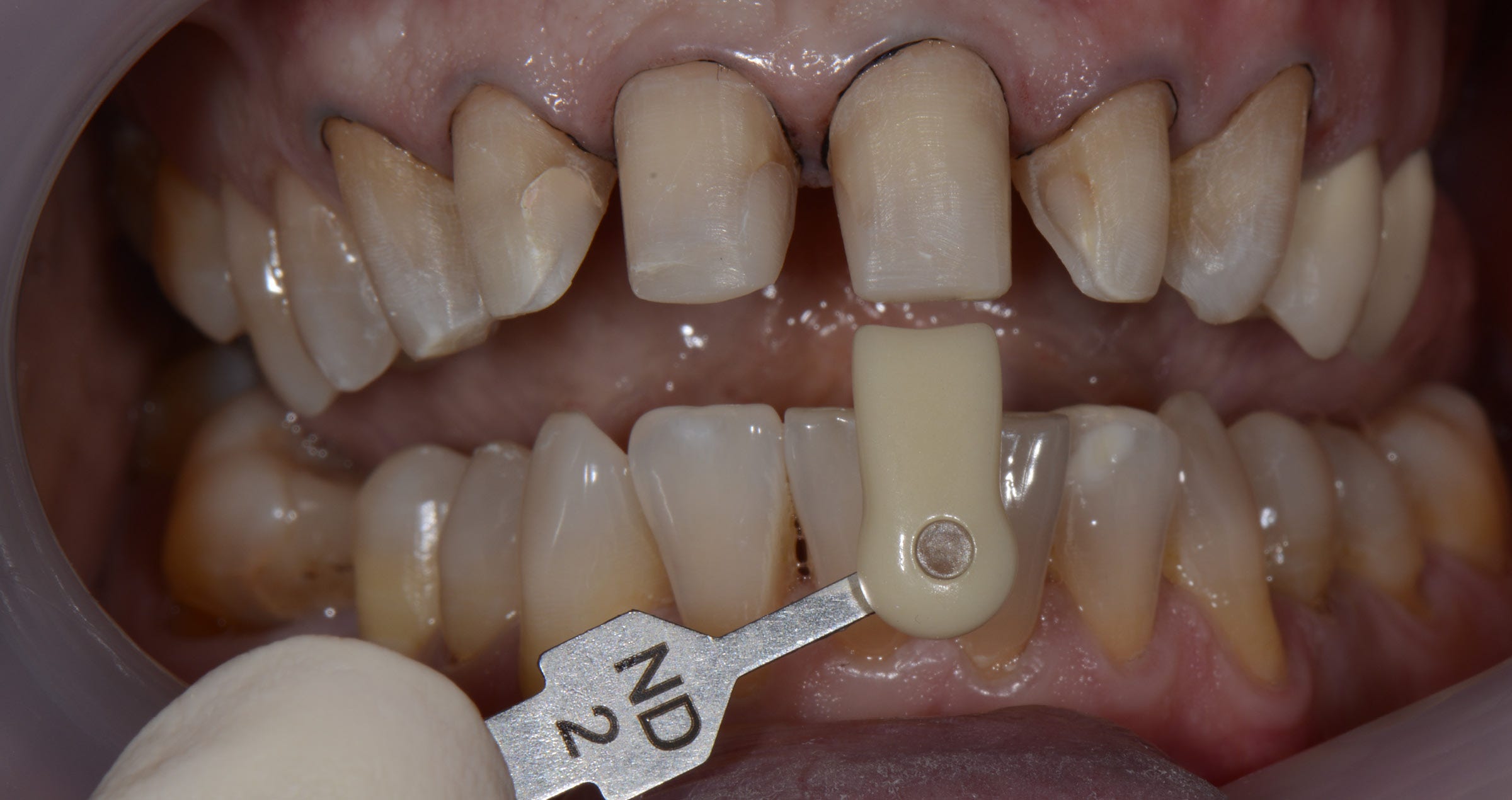
Discover a detailed walkthrough of an advanced shade reproduction technique with this comprehensive guide by DT Tomáš Forejtek. Tailored for professionals working with CERABIEN™ ZR ceramics (Kuraray Noritake Dental Inc.) and the eLAB protocol, this case study provides step-by-step insights into achieving exceptional results, from documentation to shade selection and framework design to final polish. Whether you are refining your craft or exploring new methods, this resource is a valuable addition to your toolkit.


Go for a trustworthy ceramic and metal primer

Article by Peter Schouten


Ceramic primers vary widely in composition and effectiveness, despite what their name might suggest. Most common primers include silane, but silane alone is insufficient for pre-treating all materials commonly used in indirect restorations before bonding.


Silane—typically in the form of γ-MPS—has a strong affinity for silica- or glass-based materials. While certain metals and their oxides can chemically react with silane, other components provide a stronger and more reliable bond to metals and metal oxides and should be seriously considered.


For metal(oxide) pretreatment in bonding, the MDP monomer is far more reactive than silane. The original MDP monomer, developed by Kuraray Co., Ltd. in 1981, remains the highest-quality MDP available, as confirmed by research.1


CLEARFIL™ CERAMIC PRIMER PLUS is a single-component adhesive primer that forms strong bonds with a wide range of restorative materials. This two-in-one primer incorporates the original MDP monomer, which establishes a robust bond with metals and zirconia. Simultaneously, the silane coupling agent (γ-MPS) ensures excellent adhesion to resin composites, hybrid ceramics, and glass-based ceramics such as lithium disilicate and porcelain.


ENHANCED BOTTLE DESIGN

The primer bottle is designed for effortless one-handed operation. Its unique nozzle ensures precise dispensing, minimizing the risk of contamination and spills.


PROVEN EFFECTIVENESS OF MDP-CONTAINING PRIMERS

To enhance the bonding of prosthetic materials, use a primer containing both MDP and silane. Numerous studies have demonstrated the effectiveness of this combination.


A study by Cao, Y., et al2 confirmed the superiority of CLEARFIL™ CERAMIC PRIMER PLUS (Kuraray Noritake Dental Inc.) over three other primers in improving the bond strength between zirconia and two different resin cements.

Study by Cao, Y., et al.

The effects of four primers and two cement types on the bonding strength of zirconia.

- Clearfil Ceramic Primer outperformed three other primers in improving the bond strength between zirconia and two different resin cements.

Reymus, M., et al. concluded in their study3 that before adhesive cementation of air abraded CAD/CAM resin composites, pretreatment with a specific primer, not only containing silane but also methacrylate monomers results in successfully bonded restorations. Pretreatment using an only silane containing primer results in inadequate adhesion.

Findings from Reymus, M., et al.

Bonding to new CAD/CAM resin composites: influence of air abrasion and conditioning agents as pretreatment strategy.

- Adhesive cementation of air-abraded CAD/CAM resin composites benefits from pre-treatment with a primer containing both silane and methacrylate monomers.

- Primers with only silane provided inadequate adhesion, while the combination significantly enhanced bonding success.

Four different types of CAD/CAM ceramic materials where tested in a study by Uğur, M., et al.4 Vita Mark II, IPS E.max CAD, Vita Suprinity and Vita Enamic were primed with three different primers CLEARFIL™ CERAMIC PRIMER PLUS, G-Multi Primer (GC) and Monobond S (Ivoclar Vivadent), either after hydrofluoric acid etching or no etching. It was concluded in this study that the combined effects of MDP and γ-MPTS resulted in a significant increase in the bonding strength of the resin cement to the used ceramics.

Research by Uğur, M., et al.

Effect of ceramic primers with different chemical contents on the shear bond strength of CAD/CAM ceramics with resin cement after thermal ageing.

- Four types of CAD/CAM ceramic materials (Vita Mark II, IPS e.max CAD, Vita Suprinity, and Vita Enamic) were tested with three primers: Clearfil Ceramic Primer Plus, G-Multi Primer, and Monobond S.

- Pre-treatment included hydrofluoric acid etching or no etching. Results showed that primers combining MDP and γ-MPTS substantially improved bond strength to ceramics.

Pilo, R., et al. concluded in their study5 investigating the effect of tribochemical treatment and silane reactivity on the bonding to zirconia that MDP greatly contributes to the bonding mechanism of the silane containing primers. CLEARFIL™ CERAMIC PRIMER PLUS showed to be the most reliable and effective primer in this study.

Pilo, R., et al. Study

Effect of tribochemical treatments and silane reactivity on resin bonding to zirconia.

- Examined tribochemical treatment and silane reactivity on zirconia bonding.

- MDP significantly contributed to the bonding mechanism of silane-containing primers.

- Ceramic Primer Plus was the most reliable and effective primer.

In his study6 that forms a part of his well-known dissertation, Masanao Inokoshi and others concluded that a combined mechanical/chemical pre-treatment of sintered IPS e.max ZirCAD (Ivoclar Vivadent) results in the most durable bond to zirconia. In this case when the chemical pretreatment was performed with CLEARFIL™ CERAMIC PRIMER PLUS or Monobond Plus (Ivoclar Vivadent). Scotchbond Universal (3M ESPE) and Z-PRIME Plus (Bisco), also used in this study as chemical pretreatment primers showed significant lower bond strengths.

Research by Masanao Inokoshi

Bonding effectiveness to different chemically pre-treated dental zirconia.

- Investigated mechanical/chemical pre-treatment of sintered IPS e.max ZirCAD for durable zirconia bonding.

- Clearfil Ceramic Primer (Kuraray Noritake) and Monobond Plus (Ivoclar Vivadent) yielded the most durable bonds, outperforming Scotchbond Universal (3M ESPE) and Z-PRIME Plus (Bisco), which showed significantly lower bond strengths.


OPTIMAL PRIMING FOR ADHESIVE CEMENTING

These studies underscore the critical role of MDP-containing primers in achieving reliable and durable adhesion for prosthetic materials. Products containing only silane, such as RelyX™ Ceramic Primer (3M ESPE), are less effective at creating a durable bond between resin cements or composites and ceramic- or metal-based prosthetic materials.


Image from clinical case by MDT Rondoni and Dr. Attanasio


STRAIGHTFORWARD AND EFFICIENT

The use of CLEARFIL™ CERAMIC PRIMER PLUS is straightforward: just apply it to the bonding surface, dry it, and proceed with the following treatment step. Incorporated into your process to streamline adhesion preparation and achieve reliable results!


JUST APPLY AND DRY

CLEARFIL™ CERAMIC PRIMER PLUS may be applied to any restoration surface after the required pretreatment. Pretreat the adherent surface of the restoration as indicated:


* If your laboratory already treated with a hydrofluoric acid, cleaning and activating with K-ETCHANT Syringe just before applying CLEARFIL™ CERAMIC PRIMER PLUS is recommended.

**When using with PANAVIA™ V5 or CLEARFIL™ DC CORE PLUS


Universal prosthetic primer designed for a strong bond and procedural simplicity


Author:

PETER SCHOUTEN


References

1. Yoshihara K., et al.(2015) Functional monomer impurity affects adhesive performance, Dental Materials, Volume 31, Issue 12, https://doi.org/10.1016/j.dental.2015.09.019. Pilo, R., et al. (2018). “Effect of tribochemical treatments and silane reactivity on resin bonding to zirconia.” Dent Mater 34(2): 306-316.
2. Cao, Y., et al. (2021). The effects of four primers and two cement types on the bonding strength of zirconia. Annals of Translational Medicine. 10. 10.21037/atm-21-4909.
3. Reymus, M., et al. (2019). “Bonding to new CAD/CAM resin composites: influence of air abrasion and conditioning agents as pretreatment strategy.” Clin Oral Investig 23(2): 529-538.
4. Uğur, M., et al. (2023). Effect of ceramic primers with different chemical contents on the shear bond strength of CAD/CAM ceramics with resin cement after thermal ageing. BMC Oral Health. 23. 10.1186/s12903-023-02909-z.
5. Pilo, R., et al. (2018). “Effect of tribochemical treatments and silane reactivity on resin bonding to zirconia.” Dent Mater 34(2): 306-316.
6. Inokoshi, M., et al. (2014). “Bonding effectiveness to different chemically pre-treated dental zirconia.” Clin Oral Investig 18(7): 1803-1812.


Case report by Vasilis Vasiliou

THE ART OF RESTORING SMILES: MASTERING THE CHALLENGE OF A SINGLE CENTRAL INCISOR

Restoring a single maxillary central incisor is possibly the biggest challenge a dental technician can face in everyday work. Especially when a patient is young, it is extremely important to restore her or his smile to its original beauty. Any restoration that is perceivable as such might have a negative impact on their self-confidence and quality of life even in the long term.


A STORY OF JOY AND DESPERATION

Take Ioanna, a 14-year-old girl who presented in her dental office in a state of desperation. In the hours before, she had been floating on cloud nine: Her favourite band performed in Cyprus for the first time and she had managed to buy tickets for herself and her best friend. Thrilled, they had arrived at the concert, the band started playing and the crowd danced to the music. It felt like this was going to be the best day of her life. At the time the band played its most popular song, people were delirious, jumping up and down in ecstasy. Between all the exuberant dancing and laughing, however, Ioanna suddenly was hit by a strong push. She fell, her face hitting something hard – a seat in front of her. Pain froze time and it took a few seconds before she understood what had happened: Tasting blood in her mouth, she explored her teeth with her tongue and realized that one of her central incisors had fractured.


AFFECTING THE QUALITY OF LIFE

This is one of the many touching stories we listen to every day. A fall during a concert, a push at somebody’s birthday party, a car accident: There are many incidences that can ruin a young, beautiful smile. By paying attention to the involved patients and their stories, one will come to realize how strongly some of them are affected by all this. They cover their mouths when they laugh or hold back their smiles.


Any dental technician who is committed to restoring their lost smile in the best possible way is probably aware of the impact his or her work can have and the responsibility coming with it: A Beautiful result will restore not only their smile, but also their self-confidence, will let them start laughing happily, expressing themselves comfortably and simply enjoying social interaction again (Figs. 1 to 5). Compromised outcomes, on the other hand, might have the opposite effect. Being aware of this role should be every technician’s motivation to become better day by day. Evolve for these moments, when our work brings tears of joy to our patients.


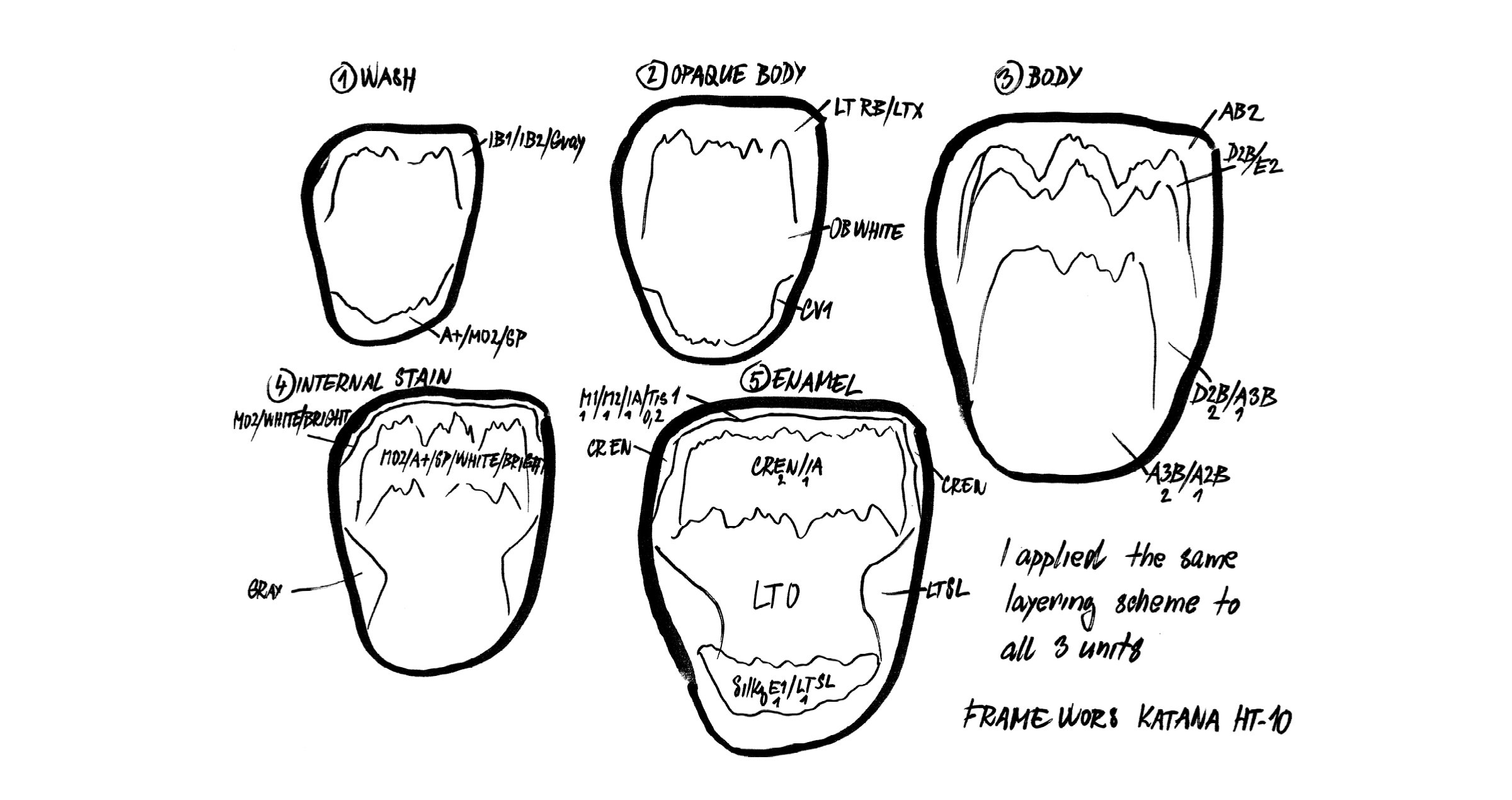
Fig. 1. Layering sketch for the restoration of a fractured central incisor in three layers: Layer one.


Fig. 2. Layering sketch for the restoration of a fractured central incisor in three layers: Layer two.


Fig. 3. Layering sketch for the restoration of a fractured central incisor in three layers: Layer three. After the first bake, small details were integrated, followed by a second bake. Finally, the restoration was finished with CERABIEN™ ZR FC Paste Stain and Glaze.


Fig. 4. Treatment outcome able to restore not only the smile, but also the self-confidence of the young girl.


Fig. 5. Immediately after cementation of the restoration, the restoration is barely identifiable, only the soft tissue needs some time for recovery.


ASPECTS TO BE CONSIDERED

But how to proceed in restoring single central incisors in the best possible way? The success of this type of restoration is hidden in the shape, which is the most difficult part. Managing to create a natural morphology is more than half the battle. The other important part is colour. The key to reproducing colour is in understanding how the utilized porcelains work. It is all about light reflection, absorption, translucency and opalescence, value and characteristic details. The more you gain experience and understand the optical properties of teeth and ceramics, the better your outcomes will be. Support is offered by a camera, a macro lens and a twin flash, which are used to capture and analyse the intraoral situation. For an initial analysis and understanding of shape and colour, I like to see the patients in my dental laboratory. Feeling the colour helps to develop the most realistic picture of what needs to be created. The key to successful realisation of the plan just developed is the use of reliable, easy-to-handle materials – in my case KATANA™ Zirconia and CERABIEN™ ZR Porcelains (both Kuraray Noritake Dental Inc.).


POSSIBLE STEPS

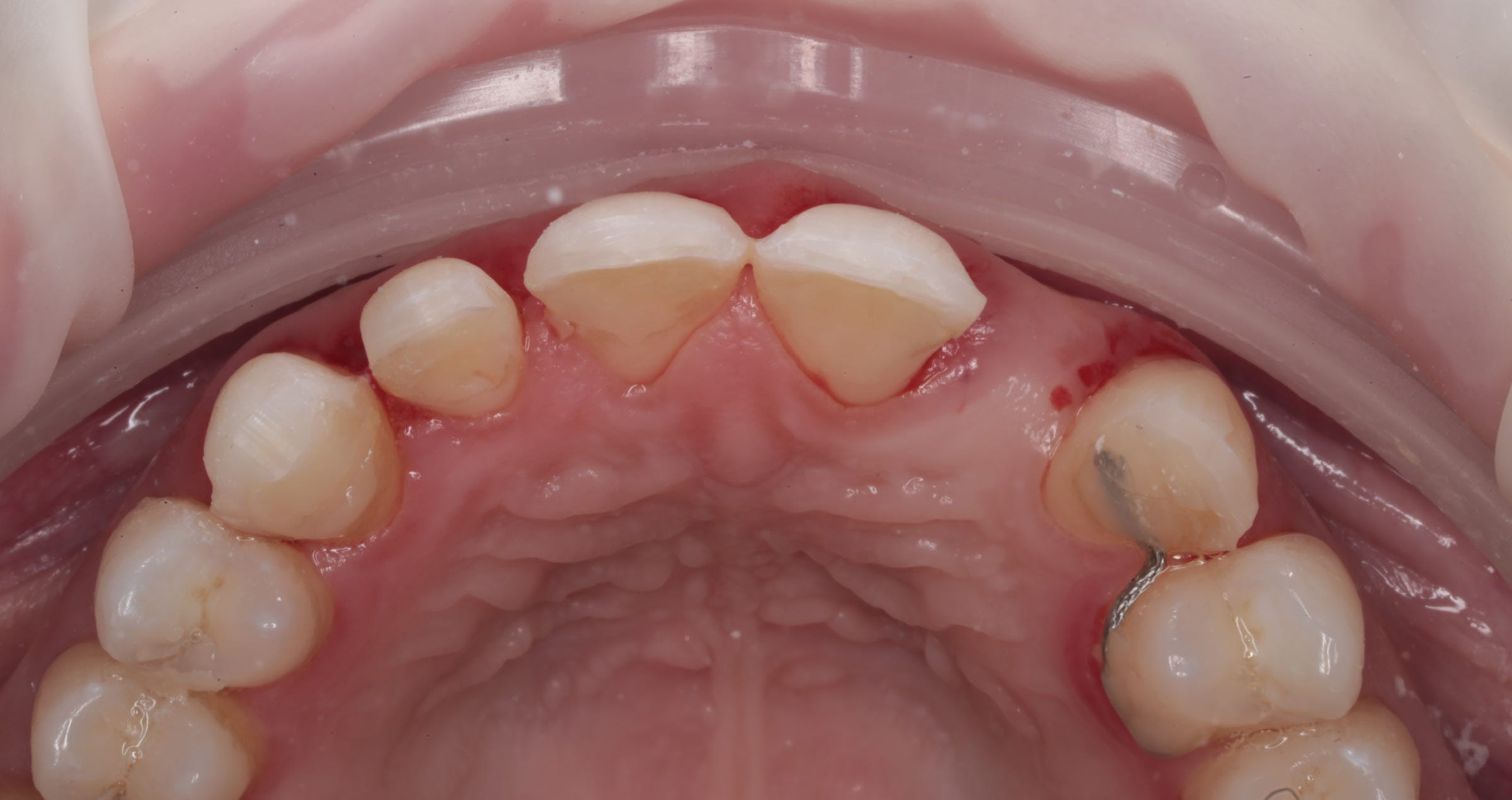
The first thing to focus on when starting to produce an anterior restoration – like in the case presented in figures 6 to 14 – is the correct value of the tooth. As soon as the framework or base is produced in the right value, you need to place what you see. Does the adjacent tooth show mamelons, traces of blue and orange? Those characteristics simply need to be observed and copied. There is no need to create something fancy. The tricky part is to use the available space reasonably. When there is plenty of space for the porcelain, it may be challenging to keep the value of the framework and avoid a greyish appearance. Depending on the die colour, age of the patient, natural surface texture and space available, an appropriate layering approach and finishing technique may be selected.


Fig. 6. Replacement of an anterior crown: Prepared tooth with severe discolouration. The adjacent central incisor has a special shape and vivid inner colour structure.


Fig. 7. Framework made of KATANA™ Zirconia ML in the shade A3. The target shade being A3.5, a quite opaque material was selected in a slightly brighter shade to achieve the required masking effect.


Fig. 8. Single-bake layering procedure: Application of CERABIEN™ ZR Opacious Body, …


Fig. 9. … Cervical Body, …


Fig. 10. … Body and Transitional Body.


Fig. 11. Incisal cut-back …


Fig. 12. … and creation of the mamelon structure.


Fig. 13. Application of Aqua Blue 1 …


Fig. 14. … followed by T Blue …


Fig. 15. … and Luster Porcelains.


Fig. 16. Halo effect created with Body.


Fig. 17. Treatment outcome. (After a first bake followed by minor adjustments, a second bake, surface texturing and glazing with CERABIEN™ ZR FC Paste Stain Clear Glaze.)


CONCLUSION

Creating a single central takes us out of our comfort zone. By paying attention, observing the adjacent teeth carefully and using materials we really understand, it is possible to meet or exceed our patients’ expectations. While specific tools like cameras and experience with the utilized materials offer support in producing predictable outcomes, my main credo is “If you want things around you to change, you must first change yourself”. For continued improvement, it is thus necessary to focus on professional growth and advancement. With the right mentors who will teach us the secrets of stratification and inspire and motivate us to continue advancing, it becomes easier to restore the smiles and self-confidence of our patients every time they need us to.


Acknowledgements

Special thanks go to the dental practitioners who treated the patients presented above – Andreas Skyllouriotis DDS, MSD, Surgically-Trained Prosthodontist, and Theo Odysseos, DDS, Diplomate, American Board of Oral Implantology / Implant Dentistry.


Dental Technician:

VASILIS VASILIOU


Vasilis Vasiliou was born in Nicosia, Cyprus, and graduated from the Technical School for Dental Technicians in Athens in 2004. He has furthered his education by attending several advanced seminars led by mentors and experts in the field, such as Ilias Psarris and Nondas Vlachopoulos.


Throughout his career, Vasilis has made significant contributions to the dental community, including presenting at various conferences in Greece and publishing articles in Greek dental magazines. Since 2020, he has been a key opinion leader for MPF Brush Company and, since 2022, a HASS Ambassador. Vasilis has been an active member of the International Team for Implantology (ITI) since 2019.


Together with his father, Vasilis runs a successful dental laboratory in Nicosia, specializing in all-ceramic and implant restorations. His extensive experience and commitment to excellence have established him as a respected professional in his field.


Digital workflows in dentistry and the future of dental care

Interview with Dr. Efe Celebi


In March 2024, Dr Ahmad Al-Hassiny, Director of the Institute of Digital Dentistry, shared his observations from LMT Lab Day 2024 in Chicago, noting that over 90 percent of U.S. dental laboratories and nearly 50 percent of dental practices have already adopted digital technologies and workflows. These advancements streamline the production of dental models, restorations across various materials, and much more.


Our company Kuraray Noritake Dental Inc. is dedicated not only to developing high-quality products and constantly adapting them to the needs of dentistry, but also to streamlining procedures in the dental laboratory and practice. Aiming to understand the current needs of dental technicians and dental practitioners around the world to provide what would really make a difference, we are in close dialogue with experts in the field. We love to listen to their stories, learn how digital technologies and artificial intelligence are already transforming dental procedures and see how we can contribute to a smooth transition – e.g. with products that support efficient workflows and great outcomes.


Lately, we had the chance to sit down with a leading figure in Turkey’s digital dentistry transformation, Dr Efe Celebi, to discuss the current landscape and future of digital innovation in dentistry. Being convinced that his experience is worth being shared with a broader audience, we have summarized the conversation.


Dr. Celebi, you’re known as a pioneer in digital dentistry. What drove you to establish companies with a strong digital focus?

As the founder of Dentgroup, Turkey’s largest Dental Service Organization (DSO), I have always believed in the power of digital dentistry. My goal was to establish digital workflows for producing indirect restorations, beginning with intraoral scanning at our practices. Initially, we sought laboratory partners in Turkey willing to make this transition with us. But at that time, none of our partner labs were prepared to take the leap, so we decided to build our own. Digitalization, after all, is simplifying work in almost every field, and dentistry is no exception. So, in 2015, we founded DentLab to provide cutting-edge digital laboratory services.


How did you go about implementing digital workflows?

From the very beginning, we aimed to digitize every aspect of our work—from production to data management and communication. Our practices already used our own practice management software, DentSoft, successfully. To connect our clinics with the lab, we developed a specialized lab module. This allowed our dental practitioners to submit every order electronically. It was a huge improvement over the old process, where forms with sketches were mailed physically, just as it had been in in the old days. Now, practitioners can select the teeth in need of treatment, specify restoration colour and design, and upload radiographs, photos, and intraoral scans with just a few clicks. Over time, we have continuously enhanced communication features between clinics and labs, adding things like delivery date notifications for orders, so patients can book their next appointment before they even leave the clinic. These tools have greatly improved coordination between our dentists and lab technicians.


How is communication organized in the software?

We created a chat-style communication area where different team members can talk, with all records saved and accessible to anyone involved in a treatment. This setup is a major improvement over phone calls, as every detail—from treatment notes to radiographs and photos—is stored and easy to reference. We even enabled practitioners to rate the products they receive, and every necessary remake is documented along with the responsible technician. This feedback system has allowed us to maintain high-quality standards, identify issues, and provide targeted training where needed.


Modern digital technologies used at DentLab to provide cutting-edge digital laboratory services.


Have you made other improvements in workflow and communication between clinics and laboratories?

DentLab initially served Dentgroup practices exclusively, but we eventually opened its services to other clinics. In this context, we set standards for incoming orders—from the required data to impression quality. If an order does not meet these standards, we reserve the right to reject it, explaining why, so the submitter can improve. We also implemented a loyalty programme and developed a special, trackable package with a QR code to prevent loss of items in transit. This innovation lets both the team in the lab and the clinic track each package’s location in real time, solving a common logistical challenge. We have even patented this unique packaging system.


Unique patented delivery box used at DentLab to prevent loss of items.


Do you provide guidance on materials and tools for clinics?

Yes, as part of our commitment to quality, we recommend specific intraoral scanners and even distribute them to customers outside our network. We also advise clinics on material choices, pre-treatment needs, and compatible resin cements for permanent placement of the produced restorations. Our protocols cover the entire restorative procedure. For aesthetic cases, we recommend starting with a smile design and using mock-ups. The mock-up evaluation lets practitioners and patients provide feedback, so the responsible lab technician can produce the final restoration with precision.


With digital workflows so well-established, do you think technicians still benefit from meeting patients face-to-face?

Not necessarily. Occasionally, a dentist may request a patient visit the lab, and we accommodate this. However, digitalization has made physical distance irrelevant, as clinics and labs can now work seamlessly from anywhere. In fact, we serve offices in Europe without any face-to-face interactions between patients and lab technicians. For patients, especially in a city like Istanbul, avoiding long travel times is a big plus, while we can still ensure high-quality outcomes.


Would you say digitalization has improved treatment quality overall?

Absolutely. There is a learning curve to digital processes, but once practitioners adjust, the quality is noticeably higher. In traditional workflows, practitioners might bend the rules, asking technicians to work with suboptimal impressions, for example. Digital systems do not allow for such shortcuts; preparation quality is clear from the scan, and impression errors can be corrected instantly before submitting to the lab. Additionally, digital scans will never shrink, distort, or tear during production, unlike physical impressions.


What are the main challenges associated with digital dentistry today?

The biggest challenge is simply taking the first step. Dentistry has been hesitant to change after decades of doing things the same way. Going digital requires an investment in both time and money. But those who make the switch find the rewards—better outcomes and more efficient procedures—well worth it.


What is next for digital dentistry?

With AI advancing quickly, the field is evolving in exciting ways. Today, we can combine digital impressions, facial scans, photographs, and 3D imaging to create a “virtual patient”. Some clinics are already using software to analyze digital data, like dental X-rays, and I predict that robot-assisted or even autonomous clinical procedures are on the horizon. Imagine robot arms taking impressions or patients scanning their own teeth with smartphones. Impression-taking procedures carried out at home already support aligner treatments in some cases. As these technologies advance, the need for dental assistants will likely decrease. The digital future for dentistry is incredibly promising, filled with tools that can transform patient care and practice efficiency on a global scale.


Example of beautiful, precisely fitting all-ceramic restorations produced at DentLab.


Kuraray Noritake Dental at the IDS 2025

INTRODUCING BRAND NEW PRODUCTS FOR SUCCESS WITH STREAMLINED, SMART SOLUTIONS

Kuraray Noritake Dental is pleased to announce that at IDS 2025 it will present a number of innovations that fundamentally improve dental procedures without compromising the quality of the result. Additionally, a number of internationally recognized experts will present their lectures and hands-on demonstrations on two separate stages: one dedicated to chair-side and the other one to lab-side topics. All of this represents not one, but a whole host of reasons to visit the stand (repeatedly).


CHAIR-SIDE NOVELTIES: UNIVERSAL EXCELLENCE

Development of the new products is rooted in a deep understanding of the demands of modern dental practice. Balancing complex procedures, tight schedules and patient needs is no easy task. That is why Kuraray Noritake´s team has been on a relentless journey to streamline, enhance and refine every step of work for decades. The vision is clear: a world where materials and tools work seamlessly in the dentist´s hands, where complexity is minimized, and where professionals are given enough time to focus on what matters most: the individual desires and needs of every single patient. Discover new additions to the UNIVERSAL EXCELLENCE family: a flowable universal composite and a new iteration of a universal bond.


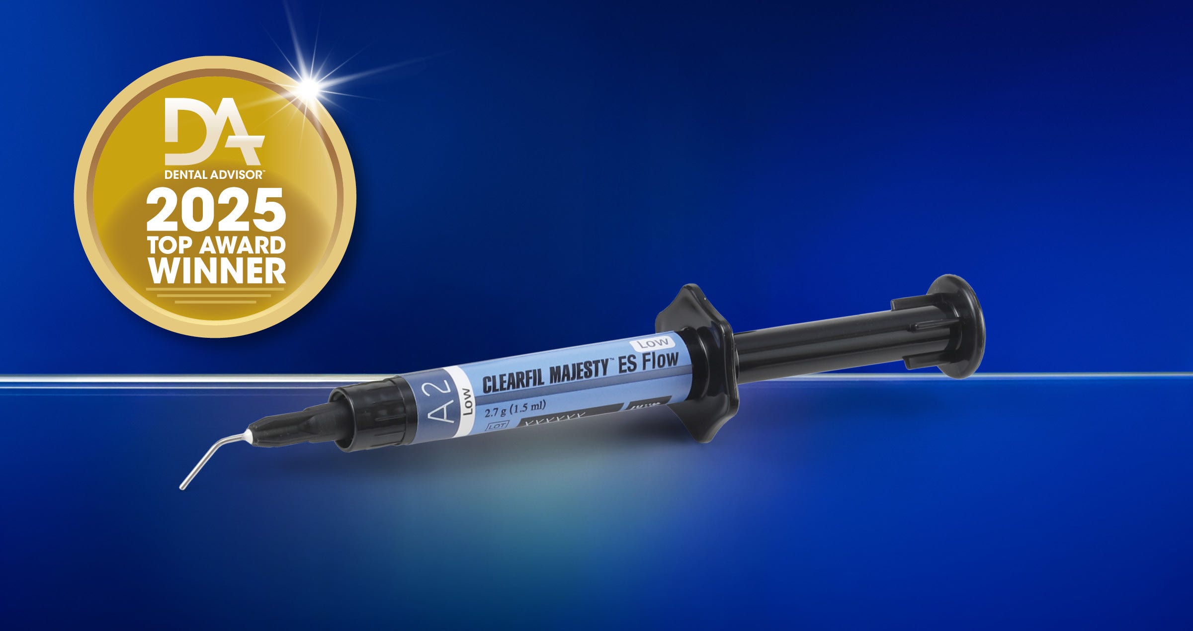
CLEARFIL MAJESTY™ ES Flow Universal represents a fundamental shift in the idea of what universal composites can achieve. By integrating high filler loading and achieving high flexural strength, CLEARFIL MAJESTY™ ES Flow Universal challenges the traditional view of flowable composites as merely temporary fixes. Instead, it positions itself as a permanent solution capable of delivering both aesthetic excellence and structural reliability. Its unique light diffusion technology allows working with just two shades, and, in addition to that, offers a choice of two flowability options for easy handling.


CLEARFIL™ Universal Bond Quick 2 is the latest evolution in dental bonding technology, offering a streamlined, efficient solution for dental practitioners. Following the success of its predecessor CLEARFIL™ Universal Bond Quick, the new version “2” offers a redefined monomer technology that delivers exceptional bonding performance with minimal effort. Kuraray Noritake´s proprietary Advanced Rapid Bond Technology combines three key monomers – the original MDP, Amide and Urethane Tetra Methacrylate. Designed to enhance workflows, this adhesive provides robust performance across a wide variety of clinical situations.


LAB-SIDE NOVELTIES

Patient-centered prosthodontic approaches are enabled by innovative restorative materials and their correct processing in the dental laboratory. No matter whether a minimally invasive procedure or the best aesthetic outcomes are desired: Kuraray Noritake Dental has the solutions. In 2025, a new “smart” finishing material is being launched, delivering both on efficiency and aesthetics.


CERABIEN™ MiLai is a set of porcelains and internal stains specifically designed for the micro-layering technique. The name “MiLai” is derived from the term “micro-layering” and the Japanese word “mirai” (which means future). The innovative product based on synthetic feldspar enables dental technicians to add the final touch to their restorations in a simple and time-saving procedure – for outstanding aesthetics right from the start. The low firing temperature of CERABIEN™ MiLai (740°C) makes it the go-to solution for the finishing of both oxide ceramic (e.g. zirconia) and silicate ceramic (e.g. lithium disilicate) restorations. With fewer lines of porcelain needed, the porcelain inventory is reduced further and fewer decisions need to be taken.


Additional information and the full programme of lectures and hands-on courses at the IDS is available online.


Performance and practicality

Case by A/Prof Alan Yap, BDS (Syd), MDSc Hons (Pros)(Syd), FAANZP


Since 1983 PANAVIA™ by Kuraray Noritake Dental Inc. has been the gold standard for dental cements throughout the world. Their latest cement, PANAVIA™ Veneer LC, sets a new standard for porcelain veneer cements through incredible performance and ease of use. The following clinical case demonstrates the use of PANAVIA™ Veneer LC.


A 31-year-old female (Fig. 1) was referred for porcelain veneers to replace lost tooth structure and to improve aesthetics. The patient exhibited moderate attrition of her anterior and bicuspid teeth (Fig. 2), the result of nocturnal bruxism and a tendency to an edge-to-edge occlusion. She had a Class I malocclusion on a Skeletal Class I tending III base with the right maxillary canine in cross-bite. The treatment plan included orthodontic treatment, porcelain veneers, and an occlusal splint.


Fig. 1


Fig. 2


Orthodontic treatment (by Dr Nour Tarraf) included full-fixed appliances with TADs and IPR of mandibular anteriors, and arch retractions to reduce protrusion (Fig. 3, post-orthodontic treatment). A preliminary digital design (Fig. 4) was performed to guide the diagnostic wax-up and a digital mock-up (Fig. 5) was utilised to verify the diagnostic wax-up prior to carrying out the intra-oral mock-up. The patient was unable to afford the restoration of the maxillary bicuspids until a later stage so the reconstruction was limited to the maxillary anterior teeth.


Fig. 3


Fig. 4


Fig. 5


Using the diagnostic wax-up, silicone keys were fabricated to guide tooth preparations. Orthodontic treatment allowed prosthetic treatment to be additive in design which meant that tooth preparations could be conservative. Labial reductions were limited to 0.3 mm and incisal reductions were performed only where needed to create an incisal butt joint for the veneer (Fig. 6). Minimal preparations allowed the veneers to be bonded almost entirely to enamel, which is important for the long-term survival of porcelain veneers (Ref 1). There was no need to significantly mask the colour of the cervical region of the tooth and non-carious cervical lesions were absent, so fine chamfer margins were prepared at equi-gingival level.


Fig. 6


Splinted provisional veneers (Fig. 7) were fabricated using bisacryl ensuring sufficient interdental space to allow hygiene access for small interdental brushes. The labial surface of the provisional veneers were glazed with a unfilled resin and cemented using the spot-etch technique, ensuring all excess flowable composite was removed prior to curing (Fig. 8). Twice daily interdental cleaning of the provisional veneers and thorough brushing of labial margins during the provisional phase maintained soft tissue health, important for the try-in and cementation of the definitive veneers.


Fig. 7


Fig. 8


A dry try-in of the definitive veneers was performed to check the fit of the veneers and a wet try-in was performed using try-in paste to assess aesthetics. The PANAVIA™ Try-in pastes accurately mimic the cement shades. Four useful shades are available (Fig. 9). The White and Brown shades are useful to correct small discrepancies in shade requiring subtle increases or decreases in shade value respectively. Conveniently the try-in pastes are the same as the PANAVIA™ V5 range of try-in pastes (excluding opaque). Following the try-in procedure the teeth were isolated using rubber dam and the floss ligature technique. KATANA™ Cleaner (Fig. 10) was used to clean the veneers prior to silanating with CLEARFIL™ CERAMIC PRIMER PLUS (Fig. 11).


Fig. 9


Fig. 10


Fig. 11


Veneers that have not been pre-etched should be etched with hydrofluoric acid prior to silanization. The use of the ProsMate™ Baton allows the cleaning, etching and silanization of all veneers simultaneously (Fig. 12). The veneers are arranged systematically on the ProsMate™ Tray ready for the cementation procedure (Fig. 13). Tooth surfaces were pre-treated with phosphoric acid (K-ETCHANT Syringe) and PANAVIA™ V5 Tooth Primer (Fig. 14).


Fig. 12


Fig. 13


Fig. 14


The newly designed cement applicator tip reduces air bubbles and the wide 16-gauge tip (Fig. 15) allows light and easy control of cement extrusion while also providing efficient wide coverage during application. PANAVIA™ Veneer LC has excellent handling because of its ideal paste consistency. It is non-sticky and its viscosity prevents the cement from flowing beyond the veneer margins until the veneer is seated. It is not runny or stringy. Furthermore its thixotropic properties results in lower film thickness during seating of the veneer. These excellent handling properties are due to the development of new filler technology which consists of spherical silica and nano cluster fillers (Fig. 16). The “touch-cure” mechanism of PANAVIA™ V5 Tooth Primer importantly seals the bonding interface while the extended working time and stability of the cement under ambient light allows the simultaneous cementation of multiple veneers. In this case all six lithium disilicate veneers (technical work by Yugo Hatai) were cemented simultaneously with PANAVIA™ Veneer LC Paste (Clear).


Fig. 15


Fig. 16


Tack-curing each veneer for one second allowed smooth and easy bulk removal of excess cement with an explorer (Fig. 17). Remaining excess of uncured paste was removed with brushes. Final curing was performed by light curing lingual and labial surfaces.


Fig. 17


The optical characteristics of PANAVIA™ Veneer LC, use of fine chamfer margins, and well-fitting translucent restorations produces a gradual and smooth transition of colour from tooth to veneer where margins disappear and soft tissues respond in a healthy way (Fig. 18). The color stability, excellent abrasion resistance and high gloss durability of PANAVIA™ Veneer LC preserves integrity and aesthetics at the margins over the long term. The extraordinary bond strength of PANAVIA™ products, so familiar to our profession over the last 20 years, is still second to none (Fig. 19).


Fig. 18


Fig. 19


“KATANA” is a registered trademark or trademark of NORITAKE CO., LIMITED - “PANAVIA” and “CLEARFIL” are registered trademarks or trademarks of KURARAY CO., LTD.


References

1. Layton DM, Walton TR. The up to 21-year clinical outcome and survival of feldspathic porcelain veneers: accounting for clustering. Int J Prosthodont. 2012 Nov-Dec; 25(6):604-12. PMID: 23101040.


Flowable injection technique: enkel, forutsigbar og reproduserbar

Intervju med Dr. Michał Jaczewski


Flowable injection technique er i ferd med å bli en populær teknikk for estetisk restaurering av flere tenner samtidig med flow-kompositt. Michal Jaczewski er en anerkjent instruktør som lærer tannhelsepersonell ferdighetene som trengs for å bruke teknikken med suksess. Han ble uteksaminert fra Wroclaw Medical University (Polen) i 2006 og driver egen privatpraksis i byen Legnica siden 2011. Han er grunnlegger av Biofunctional School of Occlusion, som driver opplæring innen emnet omfattende tannbehandlinger, og er lidenskaplig interessert i estetisk, digital tannbehandling. På IDS i Køln i 2023 viste han oss når, hvorfor og hvordan han bruker flowable injection på klinikken.


Kan du beskrive teknikken med få ord?


Flowable injection technique er en enkel, forutsigbar og reproduserbar metode å restaurere tenner på ved hjelp av flow-kompositt. Den er basert på en oppvoksing som det produseres en silikonindeks over. Indeksen fungerer så som form for injeksjonen av flow-kompositten. Kompositten lysherdes så gjennom den transparente silikonen. Den største fordelen med teknikken er at den krever liten eller ingen preparering av tennene. Den er en minimalt invasiv teknikk som kan brukes både av nybegynnere og av erfarne tannleger. Ved å bruke en kompositt med velbalansert opasitet i en tykkelse på 0,3 mm. og en spesiell protokoll for poleringen, er det mulig å oppnå fremragende resultater både optisk og morfologisk.


Når begynte du å bruke flowable injection technique, og hva er de viktigste indikasjonene?


Jeg begynte å bruke teknikken i 2018. Den var opprinnelig tenkt for restaurering av fortenner, men i dag brukes den med suksees for posteriore tenner, også. Slik jeg tenker, er den spesielt nyttig når formen på flere tenner skal korrigeres for å gi et penere smil, både for unge og eldre. Dette kan være tilfelle etter ortodontisk behandling. Tannbuen rettes opp og restaureres så til perfeksjon ved hjelp av flowable injection technique. Jeg bruker også teknikken for en makeover av smilet, for å bygge opp slitte tenner og for å øke bitthøyden ved total restaurering. I sistnevnte tilfelle, kan restaureringen enten være temporær eller permanent.


Hvordan starter du når du planlegger å restaurere en pasients tenner med flowable injection technique?


Den viktigste fasen, som har stor betydning for teknikkens suksess, er planleggingsfasen. Den består av dokumentasjon, avtrykkstaging og fremstilling av wax up og mock up og oså fremstilling av silikon-indeksen. Du kan sevfølgelig arbeide på den tradisjonelle måten med silikonavtrykk og konvensjonell oppvoksing, men bruk av digital teknologi i denne fasen vil forbedre arbeidsflyten betraktelig. Jeg starter vanligvis med foto- og videodokumentasjon og en scanning. Registrering av centric relasjon og okklusjon er også nødvendig. Så fremstiller jeg en virtuell oppvoksing ved hjelp av digital smile design software. På dette stadium er det viktig å ta hensyn til pasientens ansikts-karakteristika. Dette utføres best ved bruk av facial flow konseptet. Basert på den resulterende designen, kan et virtuelt behandlingsresultat vises og diskuteres med pasienten. Når resultatet er akseptert, printes modellen med wax up i ulike versjoner: den fulle wax upp modellen og en "interlip modell" hvor annenhver tann er med wax up og annenhver tann uten. Disse modellene brukes for å fremstille de nødvendige slikon-indeksene.


Digital Smile Design: Pasient med alvorlig tannslit.


Virtuell wax up vist i pasientens munn.


Modeller som er printet på grunnlag av virtuell mock-up.


Silikon-indeks fremstilt på alternerende (interlip) modell


Når, og hvorfor, lager du mere enn én silikon-indeks?


Det er spesielt nyttig å arbeide med den komplette og den alternerende indeksen når man planlegger å restaurere alle tennene i overkjeven. Å starte med den alternerende indeksen gir meg ekstra stabilitet og legger grunnlaget for et nøyaktig resultat, særlig når det gjelder den planlagte bitthøyden. I underkjeven, hvor håndteringen av indeksen og kompositten er vanskeligere pga. saliva og bløtvev som beveger seg, anbefaler jeg alltid å dele inn arbeidsfeltet i tre seksjoner- én anterior og to posteriore, og arbeide på dem separat.


Hvordan preparerer du tennene og sprøyter inn flow-kompositten?


I de fleste kasus, er alt vi trenger å rue opp emaljeoverflaten for bonding-prosedyren. Dette er vanligvis mulig ved hjelp av sandblåsing med aluminiumoksid (50 mikrometer med lavt trykk) Så etses emaljen med fosforsyre og det påføres en bonding. Silikon-indeksen er utstyrt med et injeksjonshull incisalt. Dette lages enkelt med spissen på kompositt-sprøyten ved at den trykkes gjennom materialet fra innsiden til utsiden. I posteriore del kan det være nyttig å bruke et fastere materiale og bruke to hull for hver tann på hver sin cusp -ett for injeksjon, og ett for evakuering. På en hard indeks må man bruke et diamantbor til dette. Jeg plasserer indeksen, injiserer kompositten fra bunn til topp, lysherder kort tid og fjerner indeksen. Endelig polymerisering gjøres etter at indeksen er fjernet og under et lag glycerin-gel. Når overskuddsmateriale er fjernet og den proksimale delen av restaureringen er polert, gjentas prosessen for de andre tennene, før restaureringene poleres.


Har du noen favorittprodukter for teknikken?

Til silikon-indeksen bruker jeg EXACLEAR (GC) fordi det er den mest transparente silikonen på markedet. Favorittkompositten min for flowable injection technique er CLEARFIL MAJESTY™ ES Flow  med lav viskositet (Kuraray Noritake Dental Inc.). På klinikken min og på kursene mine, har jeg hatt sjansen til å prøve ut mange ulike produkter. I denne sammenhengen har jeg funnet ut at kompositten fra Kuraray Noritake Dental Inc. har noen fordeler. Den er en moderne nano-kompositt med et bredt indikasjonsspektrum og stort fargeutvalg. Med de tre viskositetene kan den brukes i mange ulike kliniske situasjoner. Jeg begynte å bruke den for fem år siden til flowable injection technique. Low varianten er mitt førstevalg, fordi den er den mest allsidige og er egnet for både anteriore og posteriore tenner. De mest avgjørende grunnen for min beslutning om å bruke den, er den naturlige estetikken og de fremragende poleringsegenskapene. Du kan oppnå en spektakulær effekt uten spesielle ferdigheter. Som bonding foretrekker jeg CLEARFIL™ Universal Bond Quick. Den gjør arbeidsgangen enda enklere, raskere og mere forutsigbar. Til poleringen har jeg utviklet min egen protokoll.


Hvordan pusser og polerer du restaureringene dine?

Jeg starter approksimalt med polerstrips og noen ganger en proksimal sagtannet strips. For justering av form, har tre ulike diamanter og karbidbor vist seg å være verdifulle. deretter fortsetter jeg med fine eller ekstra-fine Sof-Lex skiver (3M) for konturering og puss og gummipolerere TWIST DIA™ for Composite (Kuraray Noritake Dental Inc.), som allerede de skaper en fin, naturlig overflateglans med lite anstrengelser. Så bruker jeg en geitehårs-børste med diamantpasta (Diamond excel, FGM) og til slutt bruker jeg et bomullshjul med en aluminiumoksid poleringspasta (Pasta Grigia II, anaxDENT). På denne måten er det mulig å oppnå en speilblank finish.


Syreets av emaljen med fosforsyre.


Fullstendig silikon-indeks på plass.


Applisering av CLEARFIL™ Universal Bond Quick.


Situasjon umiddelbart etter injeksjon av CLEARFIL MAJESTY™ ES Flow (Low), lysherding og fjerning av silikon-indeksen.


Proksimale justeringer med roterende instrumenter.


Hva er de største fordelene med flowable injection technique?


For pasienter og behandlere er den største fordelen at man sparer tid og penger. Mange pasienter har ikke råd til keramiske skallfasetter, og de er svært glade for å få tilbud om et alternativ av høy kvalitet som kan leveres i én enkelt seanse. Prosedyren er uten preparering og restaureringen kan enkelt repareres eller justeres i fargen hvis ønskelig, så risikoen ved denne behandlingen er svært lav. Tannlegen er gjerne klar for å ta i bruk teknikken etter å ha gjennomgått ett eneste kurs. Mens øvelse gjør mester, er resultatene gjerne imponerende allerede fra start. Det er ikke snakk om store investeringer hverken i tid eller utstyr for å begynne. Selvfølgelig kan man bruke mye tid på puss og polering, men jeg er sikker på at du vil finne den rette balansen mellom innsats og resultat.

Instrumentsett for flowable injection technique.


Har du noen anbefalinger om hvordan man skal begynne å bruke teknikken?


Først og fremst vil jeg oppmuntre alle til å forlate komfortsonen og begynne med noe nytt. For meg ble det å begynne å arbeide med flowable injection technique en skikkelig gamechanger, og jeg kan ikke tenke meg å arbeide uten igjen. Før du går i gang, vil jeg anbefale alle å gå på et kurs for å lære teorien som ligger bak et vellykket resultat, og kanskje også et arbeidskurs. 


Michał Jaczewski during his presentation at the Kuraray Noritake Dental booth in Cologne.


Kuraray's silanteknologi nøkkelen til flow-komposittene våre

Glem frustrasjonen over flow-kompositter. Finnes det en flow-kompositt som holder det den lover? Noen av flow-komposittene på markedet har utilstrekkelig styrke og estetikk, og er vanskelige å bruke. Dette kan gi inntrykk av at flow-kompositter er underlegne i forhold til pasta-kompositter. La oss forklare hvorfor CLEARFIL MAJESTY™ ES Flow skiller seg fra andre flow-kompositter på markedet og se hvordan avansert silaniseringsteknikk åpner for en ny æra innen flow-kompositter.

Hovedkomponentene i dentale kompositter inkluderer fillere og resiner som kombineres omhyggelig for å gi et anvendelig materiale som kan polymeriseres og som kan gi en holdbar dental restaurering. Det er essensielt at fillerpartiklene forblir i matrisen. For å oppnå dette, må fillerpartriklene feste seg til matrisen. Et bindemiddel (silan) letter den kjemiske adhesjonen mellom de to komponentene i kompositten. Silan er et bifunksjonelt, overflateaktivt materiale som binder til fillerpartiklene og reagerer med monomerene i matrisen under polymeriseringen.


SKIKKELIG SILANISERING 

Det kan virke enkelt, men virkeligheten er annerledes.. Når man blander filler og resin oppstår et problem. Mengden av f iller som kan tilsettes er meget begrenset. Meget raskt vil blandingen bli så hard at den blir nesten umulig å jobbe med. Dagens kompositter inneholder fillerpartikler som blir stadig mindre i størrelse. Ved et konstant volum, vil overflaten som skal dekkes med resin øke når partiklene blir mindre, og dette fører til at blandingen blir ubrukbar enda raskere. Det trengs derfor høykvalitets silaner slik at fuktekapasiteten til f illerpartiklene blir så høy som mulig. Dette gir en kompositt av høy kvalitet.


DET USYNLIGE MESTERVERKET

Ikke alle silaner og silaniseringsteknikker er like. Kuraray Noritake Dental viser et av sine mesterverk på dette området. Vi, som er ledende innen silanisering, bruker bare svært stabile spesialtilpassede silaner. Et av silanene som brukes er et spesialutviklet langkjedet silan. Long Carbon Chain Silane (LCSi). Dette silanet brukes i vår serie med flow-kompositter med høyt fillerinnhold: CLEARFIL MAJESTY™ ES Flow.

Resultatene av vår fremragende  silaniseringsteknologi:

  • Høyt fillerinnhold uten at det går på bekostning av brukervennlighet
  • Høy stabilitet på restaureringene selv etter påkjenninger
  • Høy fuktresistens
  • Holdbare restaureringer med naturtro utseende
  • Svært lav vannabsorpsjon
  • Optimale håndteringsegenskaper


Fig. 1: Blandinger av samme mengde resin og ulikt behandlede fillerpartikler. Til venstre: Ubehandlet, i midten: Konvensjonell silanisering, Til høyre: Kuraray behandlet.


Jo høyere kvalitet det er på silanet, desto bedre blir fuktbarheten til fillerpartiklene og desto mere filler kan tilføres til resinet uten at det går ut over håndteringsegenskapene . En tilleggsfordel ved å bruke vårt høykvalitets silan er at det bidrar til at fillerpartiklene ikke løsner så lett fra herdet kompositt. Våre kompositter har også svært begrenset vannopptak, og dette er også relatert til vår silanteknologi.

Kombinasjonen av fillerne, den før nevnte silanteknologien og en resin med lav viskositet, gjør oss istand til å skape en f low-kompositt med høyt fillerinnhold som fungerer som en universalkompositt.. Både flytegenskapene og den optimale brukervennligheten beholdes, samtidig som kompositten har høy styrke.

Fig. 2: Jo mere sofistikert silanteknologien er, desto mere filler kan tilsettes samme mengde resin.


FLYTEGENSKAPER

CLEARFIL MAJESTY™ ES Flow-familien tilbyr tre ulike viskositeter for å matche dine preferanser og type restaurering: High, Low eller Super Low.


STYRKE

CLEARFIL MAJESTY™ ES Flow har et fillerinnhold på opptil 75% (vekt), 59% (volum), og dette fører til fremragende (mekaniske) egenskaper slik at det er velegnet også for posteriore restaureringer.


HVA BETYR DETTE FOR DEG?

Hvilke fordeler gir vår ekspertise deg som tannlege?

Blant annet på grunn av den høye fillerandelen har restaureringer fremstilt av CLEARFIL MAJESTY™ ES Flow meget høy styrke. Tilstrekkelig styrke også for posteriore restaureringer. CLEARFIL MAJESTY™ ES Flow er faktisk sterkere enn de fleste konvensjonelle kompositter med pastakonsistens. (se diagrammet nedenfor). Gjentatt belastning påvirker ikke styrke og integritet negativt. Materialet er også motstandsdyktig mot fuktighet, og dette gjør at materialets styrke og estetikk holder seg stabile over lang tid. Fillerpartiklene i CLEARFIL MAJESTY™ ES FLOW består av både submikron- og klyngepartikler (0,18 til 3,5 mikrometer). De attraktive bruksegenskapene som f.eks. kontrollert applisering og at materialet ikke renner og siger, skyldes i stor grad den balanserte sammensetningen av f illere.

Typen fillerpartikler er også i stor grad ansvarlig for den holdbare høyglansen til restaureringene som er fremstilt av CLEARFIL MAJESTY™ ES Flow. De kan poleres i løpet av noen få sekunder, og selv bare å tørke av overflaten med en bomullsrull med sprit på, gir god høyglans. Lysdiffusjonsteknologien i CLEARFIL MAJESTY™ ES Flow, som hovedsaklig skyldes nanocluster fillerne, gir fremragende fargematching og gjør at restaureringen går sømløst i ett med omgivende tannvev.


KONKLUSJON

Man kan trygt si at vår ekspertise innen silanisering og andre teknologier (som f.eks. Light Diffusion Technology), har en stor positiv effekt på kvaliteten av komposittene våre, inkludert CLEARFIL MAJESTY™ ES Flow. Denne kommer i tre ulike grader av flytbarhet og tilbyr en svært anvendelig produktserie med enkel håndtering, høy styrke, enkel polering og holdbar høyglans. Enten du ønsker å bruke den til å restaurere, reparere eller sementere, anteriort eller posteriort, eller til Stamp Technique eller Flowable Injection Technique eller en hvilken som helst annen teknikk, er dette den eneste kompositten du trenger.

Dental Zirconia Og hvorfor tannleger bør involveres i avgjørelser om protetiske materialer

Viktigheten av høykvalitets protetisk behandling

Høy kvalitet på behandlingen er trolig det viktigste elementet på veien til tilfredse pasienter. Ved hvert enkelt besøk, ønsker  pasienten å føle seg ivaretatt av kompetent tannhelsepersonell, mens tid i stolen og antall konsultasjoner bør reduseres til det nødvendige minimum. I protetisk sammenheng betyr dette at en restaurering skal passe perfekt med én gang og være holdbar, for å unngå omgjøringer og ekstra tannlegebesøk.


Men hvordan er det mulig å levere høykvalitets restaureringer med perfekt tilpasning hver gang? Blant potensielle årsaker til problemer med kvaliteten på indirekte restaureringer er feil som gjøres på klinikken eller på laboratoriet, problemer med kommunikasjonen, og ofte noe som blir oversett, bruk av zirkonium av dårlig kvalitet.


Zirkoniumrestaureringer, moderne og estetiske dentale løsninger.

For over 20 år siden kom zirkonium inn på dental-markedet som en erstatning for metall ved fremstilling av kroner og broer. Begge materialer, zirkonium og metall, ble vanligvis kombinert med et lag porselen som var brent på metallet eller zirkoniumet. I de følgende årene fokuserte flere av de ledende dentalprodusentene (som f.eks. Kuraray Noritake Dental Inc.), på materialforbedringer. Disse forbedringene transformerte gradvis materialet fra  et opakt, hvitt kjernemateriale til et keramisk materiale med et tannlignende utseende og fremragende mekaniske egenskaper. De nyeste zirkonium-variantene som finnes med ulike grader av translucens og styrke, anses av tannbehandlere over hele verden for å være det beste behandlingsalternativet for et bredt utvalg av pasienter og indikasjoner. Én grunn er at de trenger bare et tynt lag porselen eller ikke porselen i det hele tatt. En annen grunn er at de trenger liten veggtykkelse, slik at de muliggjør konservativ preparering, samtidig som de har lang levetid. Vel å merke hvis man bruker et høykvalitets materiale.


Kvalitetsforskjeller på dentalt zirkonium

Kvaliteten på zirkonium kan variere avhengig av faktorer som renheten på råmaterialet (ikke bare zirkoniumet, men også alumina, yttria, fargetilsetninger osv.), den nøyaktige kjemiske sammensetningen, kornstørrelsen og partikkelfordelingen. Hvert trinn i fremstillingsprosessen (pulverblanding pressing,pre-sintring) har innvirkning på kvaliteten på diskene (optiske og mekaniske egenskaper).


Vanlige problemer som oppstår pga. zirkonium av dårlig kvalitet.

Problemer med de optiske egenskapene til en restaurering (translucens, fargeavvik, overgang mellom lagene på multi-layered materialer), vil alltid vise seg etter sintringen på laboratoriet. Det kan bli nødvendig å lage den om igjen. Andre ganger kan feilen oppdages først ved innprøving på pasienten, og dette vil ofte føre til at pasienten blir misfornøyd. Det samme vil være tilfelle for kasus med dårlig passform grunnet f.eks. inhomogen materialstruktur. Det som er enda verre, er dårlig biokompabilitet, som dårlig overflate, svake kanter, nedsatt bøyestyrke eller nedsatt bruddstyrke. Disse problemene kan ofte bare påvises med dyrt testutstyr som normalt ikke er tilgjengelig på et tannteknisk laboratorium. Dette betyr at feil vanligvis ikke oppdages før det oppstår et klinisk problem, som f.eks gingival retraksjon, økt plakk-akkumulering, slitasje, eller en feil som gir ubehag og smerter.


Oversikt over potensielle problemer og kliniske konsekvenser for pasienter

Potensielle problemer med sub-standard zirkonium

Potensielle kliniske konsekvenser for pasientene

Begrenset biokompatibilitet

Gingival retraksjon / inflammasjon

Ikke-homogen materialstruktur

Dårlig passform på restaureringen

Sprekker i overflaten

Estetiske problemer (translucens, farge) > omgjøringer

Dårlig overflatekvalitet: porøs overflate

Økt plakkretensjon > periodontale problemer, karies

Dårlig overflatekvalitet: Ru overflate

Vanskelig å pusse og polere > slitasje på antagonist

Dårlig kantstabilitet

Kantfrakturer og sprekker > tidlig reparasjon eller omgjøring

Lav bøyestyrke

Nedsatt holdbarhet > tidlig omgjøring

Lav bruddstyrke

Frakturer/ nedsatt holdbarhet > tidlig omgjøring


Sertifisering og standardisering av dentalt zirkonium

Dette er grunnen til at spesialister har utviklet en ISO standard (ISO 6872:2015), som beskriver in vitro tester som alle produsenter må gjennomføre på dentalt zirkonium som skal anvendes i Europa og USA, for å få FDA-godkjenning og CE-merking. De beskrevne testene brukes for å måle bøyestyrke og bruddstyrke, som antagelig er de viktigste egenskapene for langtids holdbarhet av restaureringer av materialet. Alle materialer som brukes i Europa og USA må bestå disse testene.


Hvordan du skal unngå å plassere zirkonium med lav kvalitet i dine pasienters munn

Derfor burde alle som bruker disse sertifiserte zirkoniumproduktene være trygge og i stand til å minimere materialrelatert risiko. Men den økende populariteten til dentalt zirkonium har tiltrukket seg interesse fra firmaer som vil ha en bit av kaken uten å ta bryderiet med å sikre høy produktkvalitet og gjennomgå sertifisering. Usertifiserte produkter som mangler CE-merking, har en ting felles, de utgjør en risiko for bedriften din og pasientene dine.


Så hvordan er det mulig å sikre produktkvaliteten på zirkonium på klinikken? Den gode nyheten er at det er noen enkle forholdsregler. Ved å følge dem, unngår du å sette inn piratprodukter eller lavkvalitets zirkonium i dine pasienters munn.


Tre gyldne regler for å gi pasientene dine høykvalitets zirkonium-restaureringer:

  • Bestill bare produkter som er produsert innenlands eller i en region med samme standarder som dine egne: Restaureringer som produseres i dentallaboratorier i Kina, for eksempel, har lavere krav til godkjenning (derfor mangler de CE-merking),og oppfyller antagelig ikke dine forventninger.
  • Snakk med ditt (norske) dentallaboratorium om hvor de får zirkoniumen sin fra: Forviss deg om at de kjøper zirkonium fra ledende produsenter (som f.eks. Kuraray Noritake Dental Inc.) gjennom autoriserte forhandlere.
  • Unngå tilbud som er for gode til å være sanne: Lave priser kan virke fristende, men den endelige kostnaden for en behandling kan lett bli høyere enn vanlig når komplikasjoner inntreffer.


Hvordan bruk av sertifisert zirkonium kan påvirke pasientene dine over tid.

Ved å forvisse deg om at zirkoniumen du bruker på klinikken din oppfyller de høyest mulige kvalitets-standarder, bidrar du til å bygge opp pasientenes tillit og positive inntrykk over tid. Selv om den første kostnaden for høykvalitets zirkonium er noe høyere enn for dårligere varianter, kan totalkostnadene bli lavere fordi restaureringene holder lengre og man unngår omgjøringer. En fornøyd pasient  er ofte bedre til å følge opp munnhygieneregimer, og de er mer lojale. Dette gir deg bedre rykte og hjelper til å bygge praksisen din.


Undersøk mulighetene og velg produkter fra sertifiserte produsenter.

Hvis du ønsker å ta et steg videre, kan du sammenligne sertifiserte zirkoniumvarianter fra flere produsenter og oppdage forskjeller. Kuraray Noritake Dental Inc. f.eks., er den eneste produsenten som utfører hele produksjonsprosessen, inkludert produksjon av råvarer, i egen bedrift. På denne måten har selskapet kontroll over hvert eneste trinn og kan sørge for en fremragende produkt-kvalitet, uansett hvilken variant du velger. Med det tilgjengelige produktutvalget som består av KATANA™ Zirconia UTML (ultra-translucent, muylti-layered), KATANA™ Zirconia STML (Superior translucent, multi-layered) og den høytranslucente multilayered HTML PLUS og YML (med økt styrke og gradert translucens), er det mulig å dekke så å si enhver indikasjon.


Optimizing intraoral and extraoral substrates for maximal adhesive potential

Article by Dr. Clarence Tam HBSc, DDS, FIADFE, AAACD


A NOVEL MDP-BASED SURFACTANT SOLUTION

The everyday practice of adhesive restorative dentistry, whether utilizing direct or indirect restorations, is fraught with the need for ideal environmental conditions to generate an optimal prognosis. The bonding of composite resin is the foundation of direct and indirect restoratives, as it provides the link between restoration and tooth. As dentistry strives to be minimally invasive, the treatment of the bonding interface is reflected in this philosophy by the use of self-etching multi-substrate acidic monomers such as 10-methacryloyloxyldecyl dihydrogen phosphate (10-MDP). There are myriad opportunities for both intaglio and fitting surfaces to be contaminated with varying agents to the detriment of restoration prognosis.


Some of the contaminants to be considered are of course, moisture from exhalation, ambient humidity in the oral cavity, blood, saliva and artificial sources such as provisional cement during a two-stage indirect delivery technique. Moisture is an agent which is only welcomed via a controlled approach during the dentin penetration phase of priming the substrate for adhesion, however if excessive in quantity will compromise the hybridization of the interface. Blood and saliva are ubiquitous in restorative dentistry, and best controlled via the application of rubber dam as part of an absolute isolation philosophy. Contamination of the prepared surface can also occur through artificial cements or lubrication agents. Hemostatic agents such as ferric sulfate and aluminum chloride have the ability to deposit insoluble precipitates on the surface of the tooth in a manner that 33% orthophosphoric acid can only partially remove. Also considered is the particulate deposition of dentin and enamel as part of standard tooth preparation. This smear layer is residual on the dentin surface, often occludes dentinal tubules, and is an obstacle that must be overcome in order to bond to the hydroxyapatite and collagen fibrils of the surface.


Overall, the risks to adhesive compromise and at worst, adhesive failure are high. This report details the use of a novel solution for debriding both indirect restorative and tooth intaglio with a 10-MDP salt-based solution that has the flexibility to be used both extraorally and intraorally.


ENDEMIC CONTAMINANTS: MOISTURE, BLOOD AND SALIVA

Moisture is a critical component to maximize the adhesive bond strength of certain modern universal adhesives. The presence of moisture allows for increased penetration of bonding solutions into dentinal tubules and between collagen fibrils, ultimately bolstering the resilience of the hybrid layer1. During the cementation of an indirect restoration, both salivary and blood contamination of the mating surfaces have been shown to have a deleterious effect on bond strengths, with blood contamination faring the worst in all conditions2. Van Meerbeck et al reported on technique sensitivity with one-step contemporary universal adhesives3. The basis of his findings note that these adhesives require water as an ionization medium for the self-etching reaction, with the need to evaporate water from the interfacial surfaces in order to maximize bond strengths. Despite this, these interfaces are considered semi-permeable which predisposes the hybrid layer to an increased risk of hydrolytic degradation in adhesive solutions that are not 2-hydroxyethyl methacrylate-free (HEMA-free), which has a greater affinity for water.


Periera et al tested varying degrees of wetness of dentin substrate controlled with variables such as short vs. long air blasts, wet vs. dry cotton pellets, microbrush use and an intentionally over-wet surface. In all groups, the “wettest” dentin intaglio surface resulted in the lowest shear bond strength4.


The influence of saliva and blood contamination is clearly negative in situations where the bonding interface was contaminated before or after adhesive application. For saliva, this reduction is due to the deposition of salivary glycoprotein on the surface, and relative to blood, macromolecules such as fibrinogen and platelets block access to the tubules for effective bonding. Blood contamination was found consistently to be more profoundly deleterious on bond strength relative to saliva2.


In general, on smear layer-affected dentin, chlorhexidine was consistently superior to other agents such as ethanol, EDTA, aloe vera in establishing the highest shear bond strength to dentin. On dentin that had previously been etched and contaminated with blood and saliva, the agent subsequently applied that showed the highest recovery of shear bond strength was 37.5% phosphoric acid5. A study on the nanomechanical and nanoroughness of etched dentin and self-etching adhesive treated dentin both contaminated with saliva revealed that KATANA™ Cleaner was capable of restoring control values of complex modulus and nanoroughness relative to control6.


SYNTHETIC WORKFLOW CONTAMINANTS: DENTAL STONE, HEMOSTATIC AGENTS, ROOT CANAL SEALERS AND PROVISIONAL CEMENTS

A 2020 study by Marfenko et al demonstrated that salivary contamination showed significantly lower bond strengths relative to intaglio contamination by dental stone from laboratory processes. The application of a silane coupling agent to the intaglio surface has a protective effect on the bond strength7. The caveat is that lithium disilicate-based restorations are often requested pre-etched with hydrofluoric acid from the laboratory. Often, the case is returned to the clinician on the secondary or primary model. If already treated with hydrofluoric acid, the surface can now be considered recontaminated with the stone or resin model or simply skin oils from handling. The unprotected surface needs to be decontaminated in any case following the try-in procedure, which now may feature elements of dental stone, blood and saliva, not to mention hemostatic agents such as aluminum chloride and ferric sulphate. If silane coupling agents are applied prior to try-in, the question of whether the intaglio surface was truly contaminant-free after removal from the model.


Aluminum chloride is a hemostatic agent that leaves an insoluble precipitate on the surface of the dentin, that is only partially removed when treated with phosphoric acid, resulting only in a partial recovery of shear bond strength relative to control. The application of ethylene diacetyl tetrasodium acetate (EDTA) returned the bond strengths to the level of normal dentin8. The bonding of polycrystalline ceramic restorations and metal alloys is contaminated with saliva upon try-in. This can be removed via steam cleaning and air particle abrasion set at 2.5 bar for 15 seconds9. Phosphoric acid is often mistakenly applied as a cleaning agent to the intaglio surface. In polycrystalline ceramics such as tetragonal zirconia polycrystal, this is disastrous, as phosphates will bond firmly to the free sites that the 10-MDP monomer normally bonds to as part of the APC protocol of zirconia bonding, significantly compromising bond strength (Blatz, 2016)10. A study of modern surface cleaners demonstrated successful debridement of the surface using KATANA™ Cleaner for both blood and saliva-contaminated substrates11, 12.


Provisional cements are thought to have a deleterious effect on the shear bond strength of adhesively-bonded indirect ceramic restorations. Ding et al (2022) uncovered that resin-based and non-eugenol cement use in the provisional phase decreased the bond strength relative to control, whilst the use of calcium hydroxide and polycarboxylate cements exhibited acceptable metrics. Debridement of the prepared surface with air particle abrasion (APA) resulted in recovery of decreased bond values to that of control13. Equally useful was the application of Immediate Dentin Sealing (IDS)14, a technique characterized ideally by APA before adhesive bonding and the application of a resin coat, occluding both the dentin tubules as well as the oxygen inhibition layer, allowing the resin-dentin bond to mature and strengthen in the absence of stresses. This approach is effective in minimizing post-operative hypersensitivity and bacterial ingress, as well as optimizing the shear bond strength particularly when indirect ceramics are concerned15. Hardan et al found that the shear bond strength was highest when IDS was completed using a three-step etch and rinse adhesive protocol14.


Hemostatic agents used in clinical dentistry exhibit a pH of 1.1 to 3.0 and are as acidic as self-etching primers16. Chaibutyr and Kois found that dentin when contaminated with 25% aluminum chloride or 13% ferric sulphate demonstrated a significantly lower shear bond strength to dentin, which was significantly recovered using the etch-and-rinse approach17. This approach albeit successful was only able to achieve partial reversal of shear bond strength deficits relative to control, with a pre-etching application of EDTA required in order for full recovery8. KATANA™ Cleaner was found to have a positive effect on the cleaning of dentin contaminated with both aluminum chloride and ferric sulphate.


The bonding of dentin substrate contaminated with root canal sealers is a concern for the integrity of core buildups post-endodontic treatment. The use of  KATANA™ Cleaner was found to be generally superior to the ethanol test subgroup in the removal of zinc-oxide eugenol-based sealer with equal performance to 70% ethanol for the epoxy resin-based sealer18.


CLINICAL CASE DEMONSTRATION

A 35 year old ASA 1 female patient presented to the practice with multiple failing composite restorations in the second quadrant that were planned for replacement. Prior to the delivery of topical and local anaesthesia, it is common procedure in the practice to ascertain shade specifics of planned restoratives before potential dehydration can affect the optical properties of the natural tooth. Smart monochromatic composites (Fig. 1) are a class of direct restoratives that leverages the ability of its nanofiller composition and refractive index to mimic the structural color of the surrounding enamel and dentin19. This typically enables a clinician to have a simplified selection of shades on hand.


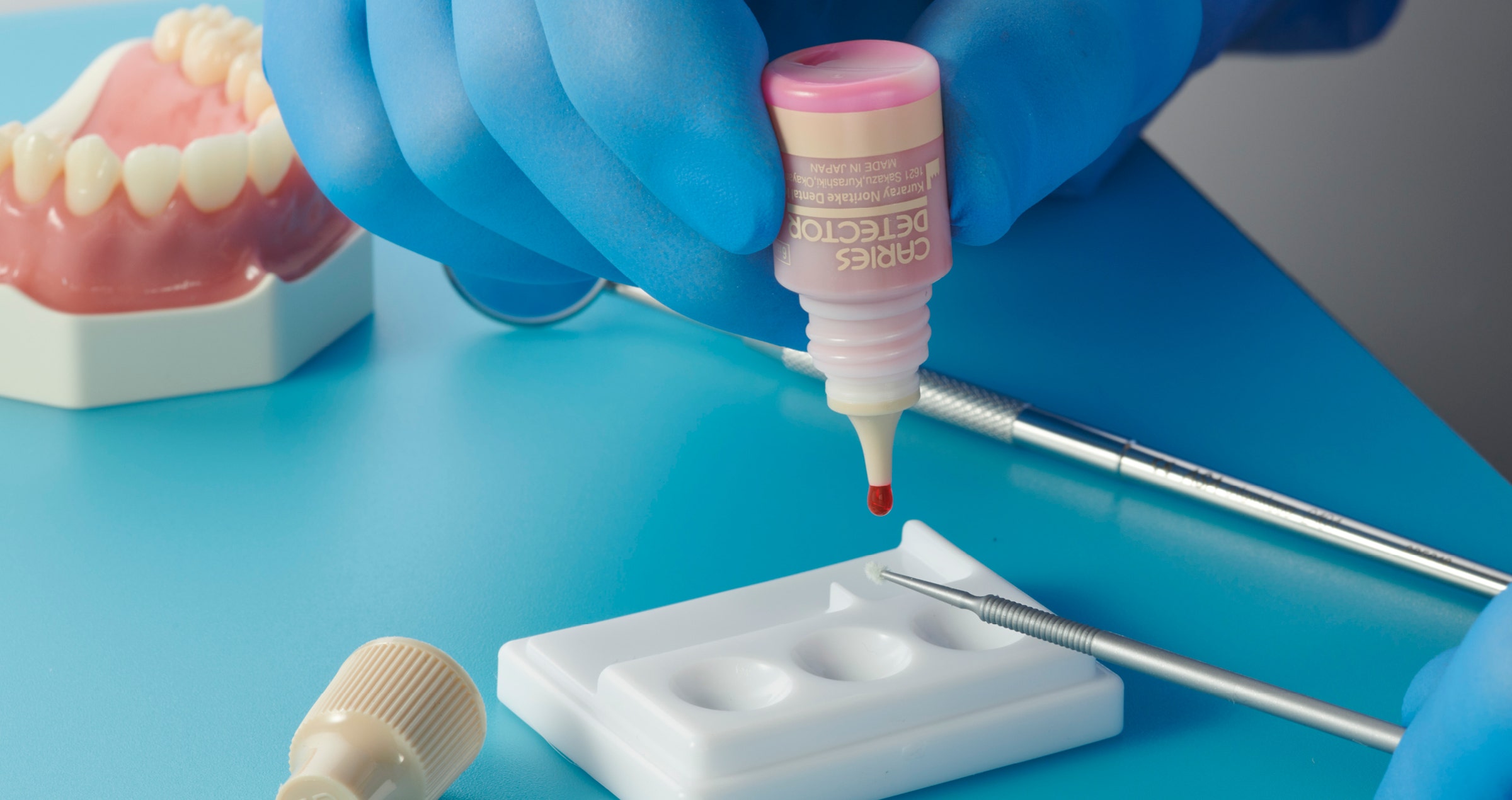
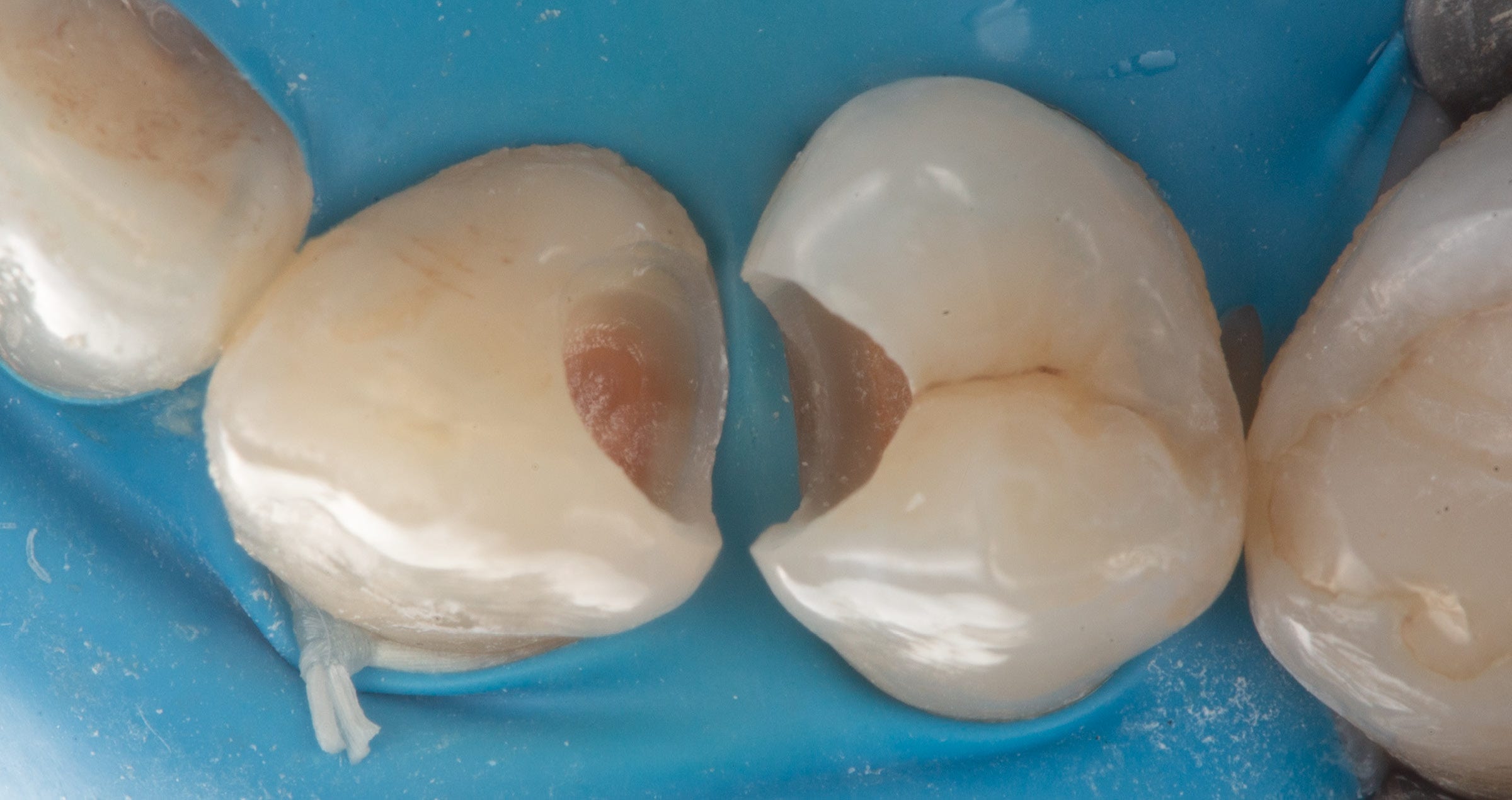
Two carpules of 2% Lignocaine with 1:100,000 epinephrine were delivered via buccal infiltration before absolute isolation was achieved using a non-latex rubber dam (Isodam HD Heavy, 4D Rubber, UK) (Fig. 2). The old restorations were excavated along with caries (Fig. 3), and the dentin structure assessed for residual decay with a detector dye (Caries Detector, Kuraray Noritake Dental Inc.). The preparation cavosurface margins were gently bevelled before surface treatment with air particle abrasion (30psi, 29 micron aluminum oxide in a 17.5% ethanol carrier, Aquacare UK) (Fig. 4). The enamel margins were etched with 33% orthophosphoric acid and rinsed (Fig. 5). The preparation surfaces were decontaminated further of any residual smear or powder residue using a MDP-based surfactant (KATANA™ Cleaner, Kuraray Noritake Dental Inc.) (Fig. 6). A single step self-etching universal adhesive was applied to the preparation as per manufacturer instructions and air thinned before light curing (Fig. 7).


Fig. 1


Fig. 2


Fig. 3


Fig. 4


Fig. 5


Fig. 6


Fig. 7


A matrix-in-matrix approach was utilized for the second bicuspid as the first step to allow for simultaneous anatomic construction of the mesial and distal marginal ridges. This technique does not require the use of a wedge as the outer circumferential Tofflemire matrix (Omnimatrix, Ultradent Products) tightens cervically around the inner anatomically-curved sectional matrix (Garrison Firm Band, Garrison Dental Solutions) allowing for a hermetic gingival seal (Fig. 8). If required, the setup may be further modified by the use of Teflon inserted between the two matrices to provide better proximofaciolingual adaptation. As a result, finishing and contour creation post-band removal is kept to a minimum. Following this, a traditional sectional matrix system may be employed to close contacts and build marginal ridges in the conventional manner (Fig. 9).


Following marginal ridge construction, the matrix assembly was removed and with the Class II lesions converted into a Class I situation, microlayering proceeded with a high flexural strength flowable liner (CLEARFIL MAJESTY™ Flow, Kuraray Noritake Dental Inc.) prior to the application of a monochromatic composite resin (CLEARFIL MAJESTY™ ES-2 Universal U shade, Kuraray Noritake Dental Inc.). The buccal cusps were constructed first as the author considers this essential to establishing restoration lobe proportions (Fig. 10). Subsequent layers were completed in a lobe-by-lobe approach to finish the occlusal anatomy (Fig. 11 and 12). The restoration was checked for occlusal functional conformativity, finished and polished to high shine (Fig. 13).


Fig. 8


Fig. 9


Fig. 10


Fig. 11


Fig. 12


Fig. 13


DISCUSSION

Dental substrates are often contaminated in both direct and indirect restorative processes. Historically, etch-and-rinse approaches have been successful for at least the partial recovery of bond strength however it is not practical in situations where selective or self-etching is the adhesive strategy. The restorative dentist in these cases can use the 10-MDP monomer in self-etching systems to target non-demineralized dentin such as CLEARFIL™ Universal Bond Quick to establish an acid base resistance zone (ABRZ) otherwise known as Super Dentin20. The presence of the operative smear layer impedes full access of the self-etching primer to the dentin substrate in some cases. In such cases, without KATANA™ Cleaner, APA is required to transform the substrate back to control bonding potential. APA as a modality is only utilized by a subset of dental practitioners often due to financial constraints or lack of technique experience. KATANA™ Cleaner thus represents a versatile tool for the decontamination and  optimization of substrate surfaces for adhesive bonding both in intraoral and extraoral applications. Its ability to re-establish ideal bonding values in areas that are not effectively reached by APA such as endodontic canal anatomy in a non-invasive manner literally cements it as a truly indispensable tool for the modern restorative dentist.


Disclaimer: Some indications are not described in the product’s Instructions for Use and are based on published research and/or the author’s experience. Before using each product, read carefully the Instructions for Use supplied with the product for full details and workflows.


Dentist:

CLARENCE TAM


References

1. Sugimura R, Tsujimoto A, Hosoya Y, Fischer NG, Barkmeier WW, Takamizawa T, Latta MA, Miyazaki M. Surface moisture influence on etch-and-rinse universal adhesive bonding. Am J Dent. 2019 Feb;32(1):33-38. PMID: 30834729.
2. Taneja S, Kumari M, Bansal S. Effect of saliva and blood contamination on the shear bond strength of fifth-, seventh-, and eighth-generation bonding agents: An in vitro study. J Conserv Dent. 2017 May-Jun;20(3):157-160. doi: 10.4103/0972-0707.218310. PMID: 29279617; PMCID: PMC5706314.
3. Van Meerbeek B, Van Landuyt K, De Munck J, Hashimoto M, Peumans M, Lambrechts P, Yoshida Y, Inoue S, Suzuki K. Technique-sensitivity of contemporary adhesives. Dent Mater J. 2005 Mar;24(1):1-13. doi: 10.4012/dmj.24.1. PMID: 15881200.
4. Pereira GD, Paulillo LA, De Goes MF, Dias CT. How wet should dentin be? Comparison of methods to remove excess water during moist bonding. J Adhes Dent. 2001 Fall;3(3):257-64. PMID: 11803713.
5. Haralur SB, Alharthi SM, Abohasel SA, Alqahtani KM. Effect of Decontamination Treatments on Micro-Shear Bond Strength between Blood-Saliva-Contaminated Post-Etched Dentin Substrate and Composite Resin. Healthcare (Basel). 2019 Nov 1;7(4):128. doi: 10.3390/healthcare7040128. PMID: 31683858; PMCID: PMC6956069.
6. Toledano M, Osorio E, Espigares J, González-Fernández JF, Osorio R. Effects of an MDP-based surface cleaner on dentin structure, morphology and nanomechanical properties. J Dent. 2023 Nov;138:104734. doi: 10.1016/ j.jdent.2023.104734. Epub 2023 Oct 2. PMID: 37793561.
7. Marfenko S, Özcan M, Attin T, Tauböck TT. Treatment of surface contamination of lithium disilicate ceramic before adhesive luting. Am J Dent. 2020 Feb;33(1):33-38. PMID: 32056413.
8. Ajami AA, Kahnamoii MA, Kimyai S, Oskoee SS, Pournaghi-Azar F, Bahari M, Firouzmandi M. Effect of three different contamination removal methods on bond strength of a self-etching adhesive to dentin contaminated with an aluminum chloride hemostatic agent. J Contemp Dent Pract. 2013 Jan 1;14(1):26-33. doi: 10.5005/jp-journals-10024-1264. PMID: 23579888.
9. Yang B, Lange-Jansen HC, Scharnberg M, Wolfart S, Ludwig K, Adelung R, Kern M. Influence of saliva contamination on zirconia ceramic bonding. Dent Mater. 2008 Apr;24(4):508-13. doi: 10.1016/j.dental.2007.04.013. Epub 2007 Aug 6. PMID: 17675146.
10. Blatz MB, Alvarez M, Sawyer K, Brindis M. How to Bond Zirconia: The APC Concept. Compend Contin Educ Dent. 2016 Oct;37(9):611-617; quiz 618. PMID: 27700128. (7)
11. Awad MM, Alhalabi F, Alzahrani KM, Almutiri M, Alqanawi F, Albdiri L, Alshehri A, Alrahlah A, Ahmed MH. 10-Methacryloyloxydecyl Dihydrogen Phosphate (10-MDP)-Containing Cleaner Improves Bond Strength to Contaminated Monolithic Zirconia: An In-Vitro Study. Materials (Basel). 2022 Jan 28;15(3):1023. doi: 10.3390/ma15031023. PMID: 35160968; PMCID: PMC8838745.
12. Tian F, Londono J, Villalobos V, Pan Y, Ho HX, Eshera R, Sidow SJ, Bergeron BE, Wang X, Tay FR. Effectiveness of different cleaning measures on the bonding of resin cement to saliva-contaminated or blood-contaminated zirconia. J Dent. 2022 May;120:104084. doi: 10.1016/j.jdent.2022.104084. Epub 2022 Mar 3. PMID: 35248674.
13. Ding J, Jin Y, Feng S, Chen H, Hou Y, Zhu S. Effect of temporary cements and their removal methods on the bond strength of indirect restoration: a systematic review and meta-analysis. Clin Oral Investig. 2023 Jan;27(1):1530. doi: 10.1007/s00784-022-04790-6. Epub 2022 Nov 24. PMID: 36422719; PMCID: PMC9877054.
14. Hardan L, Devoto W, Bourgi R, Cuevas-Suárez CE, Lukomska-Szymanska M, Fernández-Barrera MÁ, Cornejo Ríos E, Monteiro P, Zarow M, Jakubowicz N, Mancino D, Haikel Y, Kharouf N. Immediate Dentin Sealing for Adhesive Cementation of Indirect Restorations: A Systematic Review and Meta-Analysis. Gels. 2022 Mar 11;8(3):175. doi: 10.3390/gels8030175. PMID: 35323288; PMCID: PMC8955250.
15. Samartzi TK, Papalexopoulos D, Sarafianou A, Kourtis S. Immediate Dentin Sealing: A Literature Review. Clin Cosmet Investig Dent. 2021 Jun 21;13:233-256. doi: 10.2147/CCIDE.S307939. PMID: 34188553; PMCID: PMC8232880.
16. Woody RD, Miller A, Staffanou RS. Review of the pH of hemostatic agents used in tissue displacement. J Prosthet Dent. 1993 Aug;70(2):191-2. doi: 10.1016/0022-3913(93)90018-j. PMID: 8371184.
17. Chaiyabutr Y, Kois JC. The effect of tooth-preparation cleansing protocol on the bond strength of self-adhesive resin cement to dentin contaminated with a hemostatic agent. Oper Dent. 2011 Jan-Feb;36(1):18-26. doi: 10.2341/09-308-LR1. Epub 2011 Feb 21. PMID: 21488725.
18. Tian F, Jett K, Flaugher R, Arora S, Bergeron B, Shen Y, Tay F. Effects of dentine surface cleaning on bonding of a self-etch adhesive to root canal sealer-contaminated dentine. J Dent. 2021 Sep;112:103766. doi: 10.1016/j.jdent.2021.103766. Epub 2021 Aug 5. PMID: 34363888.
19. Ahmed MA, Jouhar R, Khurshid Z. Smart Monochromatic Composite: A Literature Review. Int J Dent. 2022 Nov 8;2022:2445394. doi: 10.1155/2022/2445394. PMID: 36398065; PMCID: PMC9666026.
20. Nikaido T, Weerasinghe DD, Waidyasekera K, Inoue G, Foxton RM, Tagami J. Assessment of the nanostructure of acid-base resistant zone by the application of all-in-one adhesive systems: Super dentin formation. Biomed Mater Eng. 2009;19(2-3):163-71. doi: 10.3233/BME-2009-0576. PMID: 19581710.


Abonner på vårt nyhetsbrev
Gjør som tusenvis av andre som arbeider med tannhelse og motta gratis informasjon som kan hjelpe deg i din karriere. Vi vil ikke sende ut spam eller dele din emailadresse med andre.