A GUIDE TO SUCCESSFUL ZIRCONIA BONDING


Unlock the power of zirconia: perfect for adhesive cementation, the ideal material for a wide range of indications, and essential in minimal invasive dentistry. Time to trust zirconia bonding!

This article demystifies zirconia bonding, providing clear, practical steps to ensure long-term functionality and patient satisfaction, all based on scientific research. Master the three adhesion pillars: mechanical retention, chemical activation, and wetting capacity. Discover how to successfully prepare zirconia surfaces, avoid pitfalls like misapplying silica coating and silane, and choose proven bonding systems for optimal results. Optimise retention even with minimal tooth preparation and achieve reliable zirconia restorations. Say goodbye to doubts and hello to successful zirconia bonding!


Factors influencing retention

Loss of retention due to de-cementation or debonding is a common cause of dental prostheses' failure.  First, let’s have a look at how to cope with the three main factors significantly influencing retention: tooth preparation, restoration pre-treatment, and cement type/bonding.

Tooth preparation

The abutment tooth's height, angle, and surface texture must be considered to achieve sufficient retention and resistance from the preparation. The retention form counteracts tensile stresses, whereas the resistance counteracts shear stresses 4. With the proper preparation, a restoration resists dislodgement and subsequent loss.

Full coverage restorations

To achieve sufficient retention and resistance for full-coverage crowns, the tooth abutment should be at least 4 mm high, and the convergence angle should range from 6 to 12 degrees with a maximum of 15 degrees 1, 5-8.

Source; Conventional cementation or adhesive luting - A guideline, Dr. A. Elsayed, Prof. Dr Florian Beuer 


Adhering to the tooth preparation guidelines is crucial for full-coverage restorations (e.g., crowns, and FDPs). These practical guidelines are designed to achieve the required retention and resistance to make conventional luting possible. However, optimal retention and resistance are, in reality, hard to achieve. An unwanted amount of sound tooth substance often should be removed to achieve a highly retentive preparation. Moreover, several studies2,3 show that, in daily practice, the preparation angle often exceeds 15 degrees.

Minimal-invasive restorations

Minimal-invasive restorations, such as single retainer FDPs, veneers, table-tops and inlay-retained FDPs, are based on a non- or low-retentive preparation form. In this case, retention shifts from (macro-)mechanical to micro-mechanical and chemical, necessitating the use of adhesive techniques 9-11. Even though the preparations for minimal-invasive restorations largely lack mechanical retention, the long-term success of these types of restorations is well-documented when using a suitable resin cement (e.g. PANAVIA™, Kuraray Noritake Dental, Japan), including a proper pre-treatment and bonding procedure 10, 11.

In high-retentive situations, conventional luting is acceptable for full-coverage restorations*. In all other cases, choosing a resin cement is a better solution. With proper tooth preparation (e.g., shaping, (self-)etching, abrasion) and the right adhesive resin cement system, a non-retentive preparation form provides a reliable basis using mainly chemical retention and micro-mechanical retention instead of macro-mechanical retention.

*Please review the articles available regarding the debate over whether to use a conventional cementation procedure, adhesive cementing, or selective adhesive luting


Restoration pre-treatment

Zirconia is densely sintered and does not contain a glass phase. Therefore, it cannot be etched with hydrofluoric acid to create a micro-retentive etching pattern. In addition, silanes cannot effectively promote zirconia bonding. Several studies have shown that air abrasion with 50-µm alumina at a reduced pressure of 0.5 bar (0.05 MPa; 7 psi) will create a sufficient micro-retentive pattern12 and greatly enhances the wetting capacity.

In addition to air abrasion, chemical coupling agents such as bifunctional phosphate resin monomers are used on air-abraded zirconia. Bonding with phosphate monomer-containing adhesive resin systems gives very reliable results27,28. The use of phosphate monomer-based resin cement systems (e.g., Panavia [Kuraray Noritake Dental, Tokyo, Japan]) and/or phosphate monomer primers, such as CLEARFIL CERAMIC Primer Plus (Kuraray Noritake Dental, Tokyo, Japan)  on freshly air-abraded zirconia, offer the most reliable bonding methods today 13,27,28. We therefor consider MDP-based composite resin cements the material choice for our bonding procedure. However, it must be stressed that contamination of the air-abraded zirconia with saliva, phosphoric acid or other contaminants will limit the formation of chemical bonds and, therefore, must be avoided.

Avoiding contamination

For optimal moisture control, absolute isolation of the working field is crucial. Minimising  the risk of contamination, avoiding exposure to oral fluids. Before restoration placement, a thorough cleaning of the abutment tooth is essential. Following trial placement, a meticulous recleaning step is recommended to remove any potential introduced contamination. KATANA Cleaner (Kuraray Noritake Dental, Tokyo, Japan) is an ideal choice due to its unique properties. Its slightly acidic pH of 4.5 allows for effective cleaning intraoral and extraoral adhesion surfaces. Additionally, the incorporation of MDP monomer technology makes it highly efficient. The MDP salt in this product effectively bonds with contaminants, breaks them down and results in easy removal by water rinsing.


Cement type/bonding

After pre-treatment of surfaces to optimise the , it is important to understand that the properties of highly translucent zirconia differ highly from those of earlier generation zirconia. Early-generation zirconium oxides, including 3 mol% yttrium oxide (3Y-TZP), are high in strength and low in translucency. With the increase in yttria, creating 4-5 mol% yttria, or higher, zirconium oxides, the number of cubic crystals increases, resulting in higher translucency but leading to a reduction in strength. Therefore, attention must be paid to zirconia type, material thickness, restoration type, and application area. These factors may influence the choice of cement based on the adhesive properties demanded for lasting restorations and high aesthetic outcomes.


PANAVIA V5

For a resin cement system to deliver a strong bond, it is not always enough to have it contain an appropriate adhesive monomer. It is necessary for that adhesive monomer to be polymerised effectively under different circumstances. The PANAVIA™ V5 system contains an innovative “ternary catalytic system” consisting of a highly stable peroxide, a non-amine reducing agent* and a highly active polymerisation accelerator. Since this catalytic system is amine-free, the hardened cement has unsurpassed colour stability. In addition, the highly active polymerisation accelerator, one of the components in PANAVIA™ V5 Tooth Primer, is not only an excellent reducer that promotes polymerisation effectively, but it is also capable of coexisting with the (in this product) acidic MDP. This makes it possible to create a single-bottle self-etching primer. This accelerator is also responsible for the so-called touch-cure reaction when it comes into contact with the paste. Resulting in the sealing of the dentin interface and, at the same time, allowing the paste to set even in situations where light curing is limited.

*PANAVIA™ V5 Tooth Primer applied and left for 20 seconds, followed by air drying.

The second primer in the PANAVIA V5 system is CLEARFIL™ CERAMIC PRIMER PLUS, which incorporates Kuraray Noritake Dental’s original MDP and a silane. This product is used to prime zirconia but is also an excellent choice for priming silica-based ceramics, composites, and metals.


CLEARFIL™ CERAMIC PRIMER PLUS, which contains the original MDP, applied and dried.


The PANAVIA™ V5 full adhesive resin cement system consists of all three above-mentioned components, always used in the same way, independent of the material, for a straightforward procedure to ensure reliable bonding. The PANAVIATM V5 systems offer try-in pastes to visualise the final results before final cementing and confirm the appropriate shade of the resin cement to be used.


PANAVIA VENEER LC


Offering a flexible workflow and high bondability of thin, translucent restorations like veneers but also inlays and onlays, PANAVIA™ Veneer LC was designed. It is a light-curing resin cement system allowing a long working time of 200 seconds under ambient light*. This allows multiple veneers to be placed simultaneously without racing against the setting. The final light-curing can be started anytime after positioning the provisions. The PANAVIA™ Veneer LC cementing system includes PANAVIA™ Tooth Primer and CLEARFIL CERAMIC PRIMER Plus as primers to chemically interact with the adhesive surfaces.


PANAVIA™ Veneer LC Paste applied and the laminate veneer seated. In this case six veneers were simultaneously placed during one session.

Unpolymerized excess paste removed with a brush. PANAVIA™ Veneer LC Paste is a light-cured type rein cement, designed to provide sufficient working time.

This photo shows the results after the final light curing. Since the excess cement was easily removed, there were almost no cement residues.


PANAVIA SA CEMENT Universal

Still, clinicians seek efficiency and effectiveness in everyday practice by using a straightforward but durable resin cement solution. PANAVIA™ SA Cement Universal is developed to offer this ease-of-use property without losing focus on bonding properties.  PANAVIA™ SA Cement Universal is developed with the original MDP monomer in the hydrophilic paste compartment, allowing for chemical reactiveness with zirconia and tooth structure. The other compartment contains the hydrophobic paste, to which a unique silane coupling agent, LCSi monomer, is added, which allows the cement to deliver a strong and durable chemical bond to silica-based materials like porcelain, lithium disilicate and composite resin*. Furthermore, PANAVIA™ SA Cement Universal is less moisture sensitive than full adhesive resin cement systems. This also makes it the ideal cement in situations where rubberdam isolation is difficult.

*The product is available in both auto mix and hand mix options.

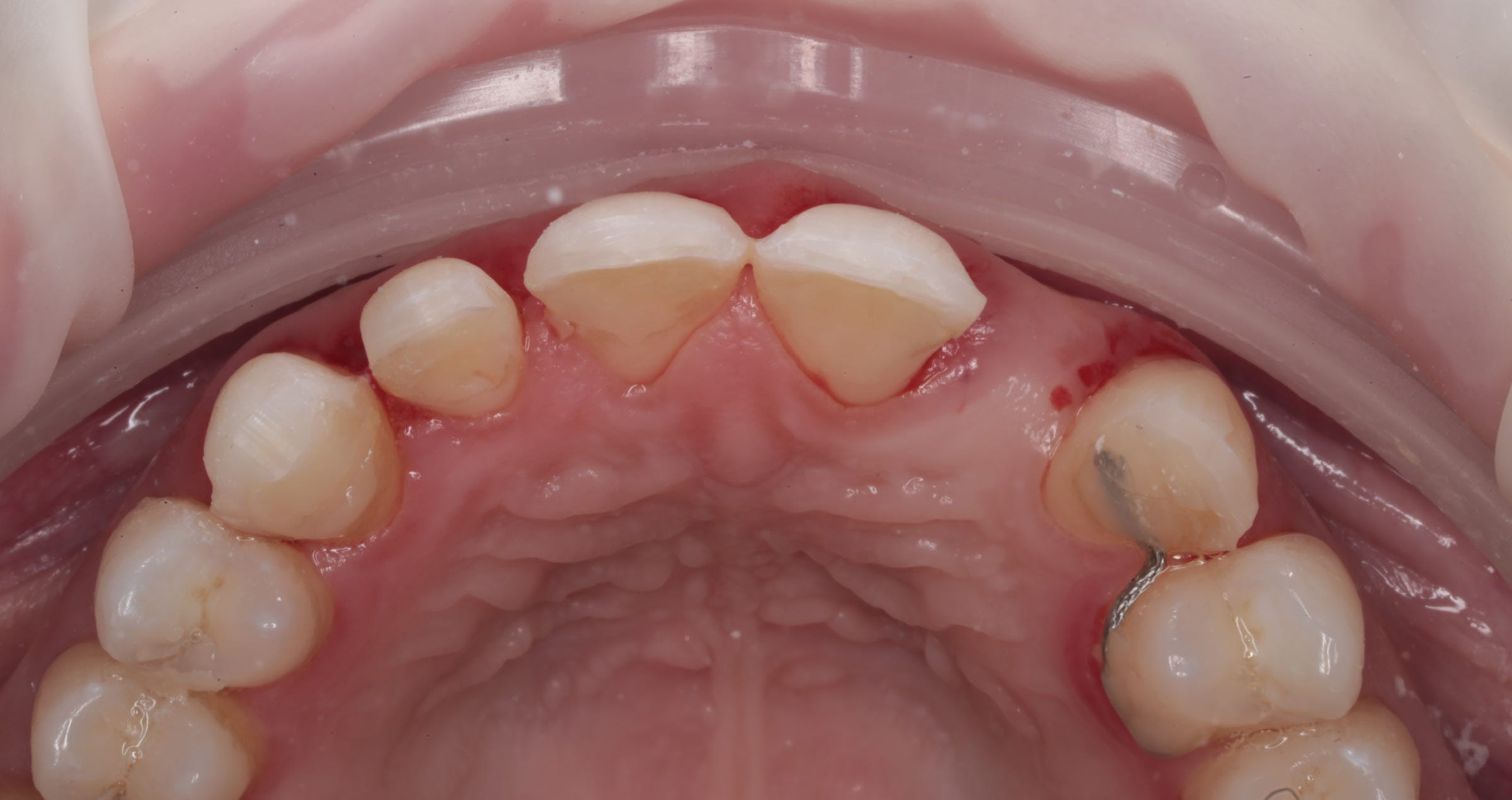
*Old PFM bridge (shown here) removed, and existing preparations modified to accommodate a 3-unit KATANA™ Zirconia bridge. The upper right canine was prepared to receive a single-unit KATANA™ crown.

Before

After. Seating & Final Smile. PANAVIA™ SA Cement Universal and CLEARFIL™ Universal Bond Quick were used for cementation and bonding. “I love the ease of use and clean-up with PANAVIA™ SA Cement Universal, and its MDP monomer creates a strong chemical bond to the tooth structure and zirconia. CLEARFIL™ Universal Bond Quick has a quick technique without reducing bond strengths, releases fluoride and has a low film thickness. I simply rub CLEARFIL™ Universal Bond Quick into the tooth for a few seconds and air dry. There is no need to light-cure, since it cures very well with PANAVIA™ SA Cement Universal. The patient was very happy with the results. She loved that she no longer saw metal margins, and her smile was much more uniform and lifelike.” Dr. Kristine Aadland


*Images are a part of a case by Dr. Kristine Aadland; 3-Unit anterior maxillary


Bonding to zirconia in three steps

Over the last century, the popularity of highly translucent zirconia has skyrocketed due to its excellent properties and wide range of anterior and posterior clinical applications. Because zirconium oxide prostheses are, if processed correctly, antagonist-friendly and easy (and relatively inexpensive) to fabricate, the material keeps gaining popularity in dentistry.

Several steps need to be taken into account for reliable and durable bonding. Years of research on achieving high and long-term bond strength to zirconia have concluded into three practical steps, summarised as the APC concept13 as a reliable procedure guideline.

APC-Step A

Zirconia should be air-particle abraded (APC-Step A) with alumina or silica-coated alumina particles; the sandblasting or micro-etching procedure. Air abrasion with a chairside micro-etcher using aluminium oxide particles (size: up to 50 μm) at a low pressure of 0,5 bar (0.05 – 0.25 MPa) is sufficient.14,18,25-27

APC-Step P

The subsequent step includes applying a special ceramic primer (APC-Step P), which typically contains specially designed adhesive phosphate monomers, onto the zirconia adhesive surfaces.29,30 The MDP monomer has been shown to be particularly effective at bonding to metal oxides like zirconium oxide.

APC-Step C

Dual- or self-cure resin cement systems should be used to reach an adequate C=C conversion rate underneath the zirconia restoration since the lack of translucency in zirconia reduces light transmission.13 However, in cases where high-translucent zirconia (HTZr02) is used, the zirconia transmits light so that the shade of composite or resin cement might influence the final appearance of such restorations. It is, thereforebased on the individual situation and shade of the abutment tooth.

The APC zirconia-bonding concept is not limited to intra-oral situations and can also be applied in the laboratory for implant reconstructions that include cemented zirconia components.

Conclusion

Rapid developments in high-quality translucent zirconia have made the utility and reliability of adhesive cementing systems even more crucial. This applies to fully opaque restorations but also minimally invasive and ultra-translucent restorations of low thickness. In all cases, the longevity of the bonding and, thus, the provision directly affects patient  satisfaction. By taking into account the three primary parameters we have discussed in this article and following the predictable APC protocol, you will successfully realise durable bonded zirconia restorations from now on.


References

  1. Ladha K, Verma M. Conventional and contemporary luting cements: an overview. J Indian Prosthodont Soc. 2010;10(2):79-88.

  2. Nam, Y., Eo, M.Y. & Kim, S.M. Development of a dental handpiece angle correction device. BioMed Eng OnLine17, 173 (2018). https://doi.org/10.1186/s12938-018-0606-1
  1. Florian BEUER, Daniel EDELHOFF, Wolfgang GERNET, Michael NAUMANN, Effect of preparation angles on the precision of zirconia crown copings fabricated by CAD/CAM system, Dental Materials Journal, 2008, Volume 27, Issue 6, Pages 814-820
  1. Muruppel AM, Thomas J, Saratchandran S, Nair D, Gladstone S, Rajeev MM. Assessment of Retention and Resistance Form of Tooth Preparations for All Ceramic Restorations using Digital Imaging Technique. J Contemp Dent Pract. 2018;19(2):143-9.

  2. Edelhoff D, Özcan M. To what extent does the longevity of fixed dental prostheses depend on the function of the cement? Working Group 4 materials: cementation. Clin Oral Implants Res. 2007;18 Suppl 3:193-204.

  3. Güth JF, Stawarczyk B, Edelhoff D, Liebermann A. Zirconia and its novel compositions: What do clinicians need to know? Quintessence Int. 2019;50(7):512-20.

  4. Smith CT, Gary JJ, Conkin JE, Franks HL. Effective taper criterion for the full veneer crown preparation in preclinical prosthodontics. J Prosthodont. 1999;8(3):196-200.

  5. Uy JN, Neo JC, Chan SH. The effect of tooth and foundation restoration heights on the load fatigue performance of cast crowns. J Prosthet Dent. 2010;104(5):318-24.

  6. Blatz MB, Vonderheide M, Conejo J. The Effect of Resin Bonding on Long-Term Success of High-Strength Ceramics. J Dent Res. 2018;97(2):132-9.

  7. Chaar MS, Kern M. Five-year clinical outcome of posterior zirconia ceramic inlay-retained FDPs with a modified design. J Dent. 2015;43(12):1411-5.

  8. Kern M, Passia N, Sasse M, Yazigi C. Ten-year outcome of zirconia ceramic cantilever resin-bonded fixed dental prostheses and the influence of the reasons for missing incisors. J Dent. 2017;65:51-5.
  1. Kern M, Dr Med Habil, M. BONDING TO ZIRCONIA. Jerd_40. 3DOI 10.1111/j.1708-8240.2011.00403.x VOLUME 2 3 , NUMBER 2 , 2011
  1. Blatz MB, Alvarez M, Sawyer K, Brindis M. How to Bond Zirconia: The APC Concept. Compend Contin Educ Dent. 2016 Oct;37(9):611-617; quiz 618. PMID: 27700128.
  1. Blatz M.B., Oppes S., Chiche G., et al. Influence of cementation technique on fracture strength and leakage of alumina all-ceramic crowns after cycling loading. Quintessence Int. 2008; 39(1): 23-32
  1. Burke F.J., Fleming G.J., Nathanson D., Marquis P.M. Are adhesive technologies needed to support ceramics? An assessment of the current evidence. J Adhes Dent. 2002;4(1)): 7-22
  1. Blatz M.B. Sadan A., Maltezos C., et al. In vitro durability of the resin bond to feldspathic ceramics. AM J Dent 2004;17 (3):169-172
  1. Blatz M.B., Bergler M. Clinical applications of a new self-adhesive resin cement for zirconium-oxide ceramic crowns. Compend Contin Educ Dent. 2012;33(10):776-781
  1. Maggio M., Bergler M., Kerrigan D., Blatz M.D. Treatment of maxillary lateral incisor agenesis with zirconia-based all-ceramic resin bonded fixed partial dentures: a case report. Amer J esthet Dent. 2012;2(4):226-237
  2. Ozer F., Blatz M.B., Self-etch and etch-and0rinse adhesive systems in clinical dentistry. Compend Contin Edus Dent. 2013;24 (1):12-20
  1. Kern M., Thomson V.P., Bonding to glass infiltrated alumina ceramic: adhesive methods and their durability. J Prosthet Dent. 1995;73 (3):240-249
  1. Kern M., Wegner S.M., Bonding to zirconia ceramics: adhesion methods and their durability. Dent Mater. 1998;14(1):64-71
  1. Wegner S.M., Kern M. Long-term resin bond strength to zirconia ceramic. J Adhes Dent. 2000;2 (2):139-147
  1. Blatz M.B., Sadan A., Martin J., Lang B. In vitro evaluation of shear bond strength of resin to densely-sintered high-purity zirconium-oxide ceramics after long-term sorage and thermos cycling. J Posthet Dent. 2004;9(4):356-362
  1. Blatz M.B., Chiche G., Holst S., Sadan A. Influence of surface treatment and simulated aging on bond strength of luting agents to zirconia. Quintessence Int. 2007;38 (9):745-753
  1. Quaas A.C., Yang B., Kern M., Panavia F 2.0 bonding to contaminated zirconia ceramic after different cleaning procedures. Dent Mater. 2007;23(4):506-512
  1. Song J.Y., Park S.w., Lee K., et al. Fracture strength and microstructire of Y-TZP zirconia after different surface treatments. J Prosthet Dent. 2013;110(4):274-280
  1. Koizumi H., Nakayama D., Komine F., et al. Bonding of resin-based luting cements to zirconia with and without the use of ceramic priming agent. J adhes Dent. 2012;14(4):385-392
  1. Nakayama D., Koizumi H., Komine F., et al. Adhesive bonding of zirconia with single -liquid acidic primers and a tri-n0butylborane initiated acrylic resin. J Adhes Dent. 2010;12(4):305-310
  1. Alnassar T., Ozer F., Chiche G., Blatz M.B. Effect of different ceramic primers on shear bond strength of resin-modified glass ionomer cement to zirconia. J Adhes Sci Technol. 2016;DOI:10.1080/01694243.1184404
  1. Blatz M.B. Long-term clinical success of all-ceramic posterior restorations. Quintessence Int. 2002;33(6):415-426
  1. Mante F.K., Ozer F., Walter R., et al. The current state of adhesive dentistry: a guide for clinical practice. Compend Contin Educ Dent. 2013;34:Spec 9:2-8
  1. Ozcan M., Bernasconi M. Adhesion to zirconia used for dental restorations: a systematic review and meta-analysis. J Adhes Dent. 2015;17(1):7-26
  1. Inokoshi M., De Munck J., Minakuchi S., Van Meerbeek B. Meta-analysis of bonding effectivenss to zirconia ceramics. J Dent Res. 2014;93(4):329-334


Tripartite talk

Presented by Kuraray Noritake Dental Inc.


Highly translucent multi-layered zirconia developed by a proprietary material and manufacturing method from Japan


CURRENT STATUS AND FUTURE PROSPECTS OF ZIRCONIA RESTORATIONS


In this issue, we asked Markus B. Blatz, Professor at the University of Pennsylvania, USA, Aki Yoshida (Gnathos Dental Studio) and Naoki Hayashi (Ultimate Styles Dental Laboratory), both dental technicians active in the USA and international instructors for Kuraray Noritake Dental Inc., to give their views on zirconia restorations and their outlook for the future.


WITH THE INTRODUCTION OF ZIRCONIA, THE MAINSTREAM OF PROSTHETIC TREATMENT HAS SHIFTED FROM METAL CERAMICS1 TO ZIRCONIA CERAMICS2. WHAT CHANGES HAVE OCCURRED WITH THE INTRODUCTION OF ZIRCONIA?


Blatz: My mentor for my first Ph.D. in dental materials was in the group that developed lithium disilicate and glass-infiltrated alumina. Therefore, I have seen the evolution of dental ceramic materials, including zirconia, which is the subject of this presentation, up close and personal.


Early zirconia was white, opaque, and not as esthetic as today. However, there is no doubt that zirconia ceramics were much more esthetic than metal ceramics. At the same time, however, we often heard the opinion that bilayer zirconia ceramic restorations were problematic, and this provoked much discussion. We conducted a large study in collaboration with a Boston laboratory to compare more than 1,000 posterior porcelain-fused-to-metal crowns and 1,100 posterior porcelain-fused-to-zirconia crowns and found no difference in chipping or fracture rates after about seven years. This proves that bilayer zirconia ceramics are safe when used with the proper veneering materials and the proper sintering and cooling protocols. The fact that zirconia became established as it is today is a major change for dentistry in general.

Yoshida: I also switched from metal ceramics to zirconia ceramics, and now I don't use metal anymore. It used to take a lot of time and effort to invest and cast metal, observe it with a microscope, and fit it. Considering the recent rise in metal prices, it has also become more cost-effective. In addition, I am allergic to metal and have a skin rash every time I have a prosthetic processed, so the shift to zirconia ceramics as the mainstream prosthetic is a welcome change. Of course, the use of zirconia has also improved esthetics. The translucency of zirconia is the greatest advantage that metal does not have.


Hayashi: Yes, that's right. The big advantage of zirconia is that if the abutment is not strongly discolored, it no longer needs to be treated with an opaquer. It was not easy to control the reflection of light from the operative tooth when fabricating metal ceramics. In addition to the esthetic advantage, the prosthetic space can be thinner than that of metal ceramics.


1. Metal ceramics: Prosthetic made of metal frame with porcelain.
2. Zirconia ceramics: Prosthetic made of zirconia frame with porcelain.


THE YEAR 2023 MARKED THE 10TH ANNIVERSARY OF THE FIRST MULTI-LAYERED ZIRCONIA – KATANA™ ZIRCONIA ML. SINCE THEN, HOW DO YOU THINK HIGHLY TRANSLUCENT MULTI-LAYERED ZIRCONIA HAS REVOLUTIONIZED PROSTHETIC DEVICE MANUFACTURING?


Yoshida: I feel the ability to extend the zirconia frame to the occlusal surface and the incisal edge is the greatest advantage of using highly translucent multilayered zirconia. This allows us to provide crowns of both esthetics and strength, even for patients with para function. I have also made a zirconia Maryland bridge using highly translucent multi-layered zirconia, and it is doing very well. There are some cases where it is not possible to use zirconia, but still, it is wonderful to have a wider range of options.


Blatz: Many people still have the impression that zirconia cannot be bonded to tooth structure, but resin cement can be used to bond zirconia to tooth structure after proper pretreatment. Clinical studies of resin-bonded zirconia bridges have shown very high success after 10 or 15 years. Currently, resin bonding is recommended for very thin, highly translucent zirconia, rather than cementation. However, it should be added that this requires the dentist and technician to understand the proper bonding technique for zirconia.


In addition, Kuraray Noritake Dental's multi-layered zirconia has revolutionized monolithic zirconia without the need for veneering porcelain. However, this has also resulted in the need for dental technicians to shift to a different approach: instead of building up the restoration as with veneering ceramics, esthetic features are created on the outer surface in each case.


Maxillary 6 anterior monolithic crowns (Markus B. Blatz)


Fig. 1a and b: Initial examination.


Fig. 1c: Simulation of final prosthetic restoration.


Fig. 1d: Completed prosthetic on model (monolithic crown using KATANA™ Zirconia STML).


Fig. 1e and f: Final restoration (Dr. Julian Conejo and Sean Han, CDT).


Two cases of Maryland bridge and laminate veneers and a mandibular canine single crown implant superstructure (Aki Yoshida)


Fig. 2a and b: Case 1: A case of a congenital defect of a lateral incisor was restored with a Maryland bridge. Since the proximal and distal width of the defect was greater than the central incisor, a non-prep veneer was fabricated on the central incisor to balance the proportions. KATANA™ Zirconia STML was used for the Maryland bridge. Note the harmony between the zirconia frame extended to the incisal edge and the transparency of the laminate veneers made of Super Porcelain EX-3™ on the central incisors. This case demonstrates the characteristics of zirconia, which combines strength and esthetics.


Fig. 3a to c: Case 2: A case of a screw-retained crown restoration of an implant placed in a mandibular canine tooth. Extension of the zirconia frame from the entire lingual side to the incisal margin prevents fracture of the porcelain by the screw access hole edges and canine guides. KATANA™ Zirconia STML provides natural transparency even when zirconia is exposed at the incisal edge.


Maxillary 4 Anterior teeth implant bridge (Naoki Hayashi)


Fig. 4a to f: Implant bridge of maxillary four anterior teeth using implants placed in the maxillary bilaterallateral incisors as abutments and maxillary bilateral central incisorsaspontics. The lingual side is fully backed with zirconia and the labial side is minimally layered with CERABIEN™ ZR.


Hayashi: Indeed, the highly translucent multilayered zirconia has expanded the possibilities of monolithic crowns. For patients with high occlusal forces, monolithic crowns are suitable in terms of strength, and with the use of highly translucent multilayered zirconia, it is possible to achieve a certain level of esthetics with monolithic crowns. In fact, some patients are happy with it. However, at least in the current situation, we believe that if patients and dentists want high-end esthetics, then porcelain buildup is necessary, and monolithic crowns are only an option.


Blatz: The variety of options available is the advantage of zirconia. The dentist and the technician can work together to provide the best possible outcome for the patient.


Yoshida: In terms of options, Kuraray Noritake Dental's zirconia can be sintered in a short time (approximately 90 minutes) in addition to the normal sintering time (7 hours) using a zirconia raw material and manufacturing method developed by Kuraray Noritake Dental, which is an advantage in that it can be used for immediate restorations, remanufacturing and other unexpected situations.


FINALLY, DO YOU HAVE A MESSAGE FOR THE NEW GENERATION OF DENTISTS AND DENTAL TECHNICIANS?


Blatz: I encourage my students and colleagues to always do their best. This leads to good results, makes you happy, and makes you feel satisfied with your life. Some people only try to get rich, but just accumulating wealth is never happiness. The second is to keep an open mind. Nowadays, we are inundated with information through social media.


Some of it is very stimulating and wonderful, but there is also a lot of it that is wrong. On the other hand, there are those who believe that everything one leader says must be done. I would like to tell them, "Make sure you get your information from reliable sources, and then choose reliable information for yourself. Dentistry is changing, so let's keep an open mind. The most important thing is that the patient is ultimately satisfied with the results.


Hayashi: I would like the future generation to learn more about tooth morphology, occlusion, and fit. Color is the essence of the quality of the final prosthetic device, but we need to learn tooth morphology, occlusion, and fit before we learn color. We are all about creating a prosthetic device that will function in the patient's mouth for the long term, and that is our goal. There will be new technologies and materials in the future, but their essence will never change. I hope that you will always remember what is important in your clinical practice. This is why basic knowledge of anatomy and function is necessary.


Yoshida: New technologies and materials will continue to emerge. But human teeth will not change. The most important thing is to provide the best possible care to the patient. I hope that you will accumulate such experiences, and that when you reach the end of your life, you will be able to say that you are glad you chose this profession.


Thank you very much for the meaningful discussion today.


Source: QDT Vol.49/2024 April
The magazine may not be printed from the web and may not be forwarded
No reproduction or reprinting allowed


Dentists:

Prof. Dr. Markus B. Blatz

University of Pennsylvania
School of Dental Medicine
240 S 40th St, Philadelphia,
PA 19104, USA

Aki Yoshida, RDT

Gnathos Dental Studio
56 Colpitts Rd, Weston,
MA 02493, USA

Naoki Hayashi, RDT

Ultimate Styles
Dental Laboratory
23 Mauchly Suite 111, Irvine,
CA 92618, USA


Empower your dental lab with KATANA Zirconia YML

KATANA™ Zirconia YML offers an unmatched blend of aesthetics and mechanical properties, but also provides for cost and time efficiencies.


Recognised for its strength and density at point of manufacture, the material delivers incredible hardness in its green state. This offers the fully validated opportunity to make adjustments in morphology directly after milling.


These qualities, along with its strength and translucency once sintered, deliver the possibility to produce a wide range of high aesthetic indications. KATANA™ Zirconia YML has set a new benchmark in prosthetic dentistry.


It provides dental technicians with a material that is truly universal with no compromises required.


KATANA Zirconia YML in a Nutshell


KATANA Zirconia YML represents a pinnacle of zirconia technology. With its multi-layered structure, it offers a seamless gradation of colour, strength and translucency that mimics natural teeth, making it an ideal choice for the entire indication spectrum.


The material's unique composition allows for high-speed sintering (up to 3-unit bridges), which significantly reduces production time without sacrificing optical or mechanical properties.


Colour Gradation and Physical Properties


The colour gradation of KATANA Zirconia YML is designed to replicate the natural colour transition of human teeth, from the dentin core to the translucent enamel surface.


This combined with the material's impressive flexural strength of up to 1,100 MPa and translucency of up to 49%, enables the production of restorations that are virtually indistinguishable from natural dentition.


Applications and Advantages of KATANA Zirconia YML


KATANA™ Zirconia YML's versatility extends to a wide range of indications, including crowns, veneers, inlays, onlays, and bridges of all sizes. With its strong body and highly translucent enamel layer, it offers exactly the properties required for an unlimited indication range.


Positioning of restorations in KATANA™ Zirconia YML discs is extraordinarily easy. The reason is that the gap between the lowest flexural strength found in the enamel area and the highest flexural strength found in the lowest body layer is comparatively small. Moreover, the Body Layer 1 that is found adjacent to the enamel layer already offers a flexural strength that is higher than the 800 MPa requested for bridges with four or more units. Consequently, the material is classified as a Class 5 zirconia and users are on the safe side whenever they place their long-span restorations in the middle of the blank.

Positioning of long-span restorations in the middle of the disc.


Revolutionizing Sintering with High-Speed Capabilities


One of the groundbreaking aspects of KATANA Zirconia YML is its compatibility with high-speed sintering protocols. This capability allows dental laboratories to expedite the production process, delivering high-quality restorations in a fraction of the time traditionally required. Sintered during normal working hours at daytime, small restorations can be finished within hours, while the sintering load at night is reduced automatically. Great option not only for rush cases!


The high-speed sintering process does not compromise the material's optical or mechanical properties, maintaining its aesthetics and strength.


Recommended Finishing Techniques for Optimal Results


KATANA Zirconia YML is a beautiful and aesthetic material in its own. Therefore, when it comes to finishing, CERABIEN™ ZR FC Paste Stain is a great option.


KATANA Zirconia YML: A Testament to Innovation in Dental Materials


KATANA Zirconia YML stands at the forefront of dental material technology, offering outstanding aesthetics, strength, and efficiency. Its introduction has marked a significant advancement in the capabilities of dental technicians, allowing for the creation of restorations that truly mimic the beauty of natural teeth in a fraction of time.


As the dental industry continues to evolve, KATANA Zirconia YML remains a testament to the relentless pursuit of excellence in restorative dentistry.


For more detailed information on KATANA Zirconia YML, including technical guide, FAQs and Clinical cases, visit Kuraray Noritake Dental's YML dedicated page.


Interested in articles, user experience or clinical cases using KATANA Zirconia YML? Check the blog section of our website! 


Mathias Fernandez Y Lombardi

EU Scientific Manager
Dental Ceramics & CAD/CAM Materials
Kuraray Europe GmbH


Discover the Latest in Dental Innovation with BOND Magazine Volume 11

Welcome to the latest edition of BOND Magazine, your essential guide to cutting-edge advancements and techniques in the world of dentistry. Volume 11 is packed with insightful articles, expert interviews, and practical advice designed to enhance your dental practice or laboratory and keep you at the forefront of the profession.


In this issue, we dive into the simplicity and predictability of the flowable injection technique with Michał Jaczewski. Learn how this minimally invasive method can transform patient outcomes with minimal preparation, making it accessible for both beginners and experienced practitioners alike.


We also explore the transformative potential of universal resin cement in Prof. Lorenzo Breschi's article, which introduces a third application mode that could revolutionize how you approach luting for challenging restorations.


Meanwhile, Dr. Michael Braian shares his comprehensive guide to dental rehabilitation using digital workflows, ensuring you can leverage the latest technology for superior patient care.


This volume also includes a case reports by Dr. Jose Ignacio Zorzin - discussion on rationalizing clinical procedures with universal adhesives. These insights will help streamline your workflows, reduce chair time, and enhance patient satisfaction.


From detailed explorations of high-performance materials to interviews with leading dental laboratory professionals like Alexander Aronin and Andreas Chatzimpatzakis, BOND Magazine offers a wealth of knowledge for all dental professionals.


Click here to read. Enjoy reading, and let us inspire your journey towards excellence in dentistry.


Start Reading: BOND | VOLUME 11 | 07/2024


Previous versions:


BOND | VOLUME 10 | 10/2023

BOND | VOLUME 9 | 08/2022

BOND | VOLUME 8 | 12/2021

BOND | VOLUME 7 | 10/2020


Flowable injection technique: an innovative minimally invasive tool

By Dr. Adrien Lavenant


In many clinical situations, composite restorations offer advantages over all-ceramic restorations. The treatment is less costly, usually less invasive, and the restorations can be modified and repaired at any time. When it comes to restoring multiple teeth, however, a free-hand layering approach used to build up every tooth separately can be very time-consuming and labour-intensive. In these situations, the flowable injection technique –also referred as injection moulding – is a great alternative. It is minimally invasive, suitable as a temporary or permanent solution to issues around tooth wear and irregular shape, and time-efficient in the clinical setting.


Essential materials

While the planning phase including the creation of the wax-up can be accomplished in the traditional way or in the digital workflow and components may differ depending on the preferred way of working, two materials are essential for the implementation of the flowable injection technique: A transparent silicone and a flowable composite. The silicone is used to produce an index and transfer the planned shape of the restorations from the wax-up into the patient’s mouth. Transparency is needed to make sure that the flowable composite will be cured properly through it – an important precondition for high-quality results. The flowable composite is injected into the silicone index. While offering a great flow behaviour during injection, the selected material should also exhibit a high mechanical stability for durability. CLEARFIL MAJESTY™ ES Flow Low (Kuraray Noritake Dental Inc.) offers the desired properties. One possible way to combine the materials and implement the technique is illustrated using the following patient case.


Important step in a complex treatment

This patient came to the dental office after orthodontic treatment with a request to improve the aesthetics of her smile. In the maxillary and mandibular anterior region (incisors and canines), severe tooth wear was diagnosed, with the maxillary central incisors most severely affected (figs. 1 to 4). To restore her maxillary teeth as quickly as possible, the least invasive immediate treatment option was selected: A smile makeover with composite using the flowable injection technique. In the long term, it is planned to perform a full-mouth rehabilitation with all-ceramic restorations.


Fig. 1. Initial situation: Lateral view from the right.


Fig. 2. Initial situation: Lateral view from the left.


Fig. 3. Initial situation: Frontal view.


Fig. 4. Close-up of the severely worn central incisors.


Shade determination and index production

After an analysis of the tooth colour using two different methods (figs. 5 and 6), a digital impression was taken and a wax-up designed with dedicated CAD software. The wax-up model was then printed (fig. 7); it served as the basis for the production of the transparent silicone index (figs. 8 and 9). After complete curing of the material, injection channels were integrated at the incisal edges of each tooth to be restored.


Fig. 5. Determination of the tooth colour using a shade guide.


Fig. 6. Picture taken with a white_balance grey reference card for objective shade quantification.


Fig. 7. 3D-printed model of the upper jaw with the virtually designed wax-up.


Fig. 8. Silicone index produced over the wax-up model with injection channels at the incisal edges of each tooth.


Fig. 9. Close-up view of the index with injection channels.


Shade validation and preparations for injection

To validate the selected shade, small amounts of composite (buttons) in three different shades were applied to the untreated surface of the left central incisor and cured (fig. 10). In this way, it is possible to visualize the colour of the composites in the mouth. The selected shade was A1. The aprismatic enamel layer on the surfaces of the six maxillary anterior teeth was carefully removed with burs to create ideal bonding conditions (fig. 11). The teeth were then isolated with clear matrix strips for the implementation of the bonding protocol on the right canine and central incisor and left lateral incisor: To provide for proper interproximal separation and contacts, it is advisable to treat every other tooth and then repeat the procedure for the rest. The teeth were etched with phosphoric acid, rinsed and carefully dried before applying the selected adhesive (CLEARFIL™ SE PROTECT, Kuraray Noritake Dental Inc.) (fig. 12). For separation and protection of the adjacent teeth during composite injection, PTFE tape is a great choice (fig. 13).


Fig. 10. Composite buttons applied to the left central incisor for shade validation.


Fig. 11. Roughened tooth surfaces.


Fig. 12. Etched and bonded surfaces of the right canine, right central incisor and left lateral incisor after isolation with clear matrix strips.


Fig. 13. Protection of the adjacent teeth with PTFE tape.


Composite injection and treatment of the other teeth

After its application, the silicone index was placed in the mouth and CLEARFIL MAJESTY™ ES Flow Low in the shade A1 injected tooth by tooth through the injection channels (fig. 14). What followed was proper light curing of the composite through the index. The situation after index removal is shown in figure 15. At this stage, the excess still needed to be removed, before protecting and separating the already restored teeth with PTFE tape and repeating the procedure for the other lateral incisor, central incisor and canine.


Fig. 14. Index with injected composite.


Fig. 15. Result after index removal.


Treatment outcome

Once all the excess material was completely removed, the occlusion was checked and adjusted. Finishing and polishing was accomplished with TWIST™ DIA for Composite (Kuraray Noritake Dental Inc.). The immediate treatment outcome is shown in figures 16 to 18, while figures 19 and 20 were taken at a recall after six months.


Fig. 16. Treatment outcome: Frontal view.


Fig. 17. The new smile.


Fig. 18. Treatment outcome: Occlusal view.


Fig. 19. Appearance of the teeth…


Fig. 20. … at the six-month recall.


Conclusion

Thanks to advances in restorative materials and technological tools, it is nowadays possible to restore our patients' smiles quickly and reproducibly. The flowable injection technique is one of the most successful examples of modern aesthetic treatments using composite resin. CLEARFIL MAJESTY™ ES Flow has all the qualities needed to carry out these treatments under the right conditions.


About the Author

Dr. Adrien Lavenant obtained his degree in Dentistry from Aix-Marseille University in 2010. He pursued post-university training in periodontology, implantology, and restorative and aesthetic dentistry (Aix-Marseille University and Paris). Dr. Lavenant has been a former teaching staff member at Aix-Marseille University since 2011 and continues to teach in the postgraduate programme in restorative and aesthetic dentistry. He practices in his private clinic in Aix-en-Provence, specializing in restorative, prosthetic, and aesthetic implant dentistry. He has been a member of the international Bio-Emulation group since 2019 and shares his patient care philosophy in accordance with the principles of biomimetic dentistry.


Kiyoko Ban - A legacy in the field of dental technology

By Manabu Suzuki, Director of Dental Division, Kuraray America, Inc.


Kiyoko Ban, a prominent figure in the dental technology field, has made a lasting impact as a researcher, developer, and founder of Noritake Dental business (Fig. 1). Renowned for her contributions to dental porcelains like Noritake's CZR and EX-3, and KATANA™ Zirconia, Ms. Ban stands as a pivotal force in the global advancement of dental technology, earning her the esteemed reputation of developer and marketer within the dental technology community.


After completing her university education in Nagoya, Ms. Ban initially assisted in her family's gas station business. However, driven by a desire for a career change, she enrolled in a newly established dental technician college in Nagoya at the age of 30. Her aspiration was to enter a field where gender distinctions held no sway, offering the potential for worldwide recognition based on technical mastery.


In 1977, a college-sponsored tour to American dental laboratories ignited Ms. Ban's dream to work in the United States. However, she delved into research across various fields such as chromatology (the science of color), ceramics and metals, finding a newfound passion for research over clinical work after graduation because she was offered a "Curriculum Chief" position from the college when she graduated (Fig. 2).


Fig. 2. Ms. Ban, a curriculum chief at the Dental Technicians College, devoted her evenings to material research.


Fig. 3. In the 1990s, Ms. Ban actively engaged in promoting EX-3 through sales efforts in Italy.


Her teaching career spanned from the age of 34 to around 40, during which she pioneered porcelain training sessions for technical improvement and arranged lectures over weekends by famous speakers such as Masahiro Kuwata.


At the age of 40, she resigned teaching career and pursued her research career. The opportunity to conduct full-scale experiments led her to the discovery of a company with advanced ceramic technology "Noritake Co., Limited", renowned for its tableware. In 1986, Cusp Dental Supply, a research institute, was established by Ms. Ban in Nagoya, focusing on the development of materials for PFM crowns. The commercialization of Super Porcelain AAA (EX-3) in 1987 marked a significant milestone, addressing issues prevalent in porcelain materials of that time, such as cracks, greening, and fluorescence.


She began traveling all over Japan and around the world to sell the products she had developed and went on to develop new products that were needed by dental technicians worldwide (Fig. 3). She continued to develop new products such as CZR, CZR Press, and KATANA™, the world's first multilayer zirconia.


Ms. Ban has been actively involved in mentoring students and graduates seeking opportunities to work overseas. During summer vacations, she took students and professionals interested in working abroad to countries like Australia, Germany, and the United States. The aim was to visit dental clinics, dental technician schools, and laboratories, fostering exposure and learning in an international context.


Simultaneously, Ms. Ban delved into researching non-precious dental technology. Inspired by her exposure to the term "non-precious" during her time in the United States, she anticipated its potential in Japan. Her research presented at lectures and events highlighted the shift in the landscape as the price of gold surged, rendering precious alloys containing significant amounts of gold impractical for PFM crowns.


As the demand for their developed products grew, the need for global acceptance became apparent. In 1990, Cusp Dental Research was established in Manhattan, New York, marking Ms. Ban's foray into establishing a company overseas. Despite the unfamiliarity with legal procedures and the challenges of setting up a foreign company, Ms. Ban, driven by determination, overcame these hurdles. The establishment of the company in the United States expanded their presence internationally Fig. 4).


Fig. 4. Capturing the essence of ISC 1996 - the International Symposium on Ceramics in Orlando, FL..


Noritake Dental Supply Co., Limited was established in 1998 by the Noritake Co., Limited, which aimed to further expand its dental business. Despite the absence of a capital relationship with Noritake at the time of establishing the research laboratory, Ms. Ban played a key role in joint research efforts with Noritake. Then she was invited to this company as the position of president, owning 60% of the stock, while Noritake held 40% (Fig. 5).


Besides Noritake Dental business, she continued expansion with the establishment of a dental laboratory in Boston in 1995. Despite the challenges posed by the September 11, 2001 World Trade Center incident, they acquired their building in Boston, integrating their New York laboratory into the Boston operations.


Ms. Ban's tenure as president of Noritake Dental Supply persisted until 2009, but organizational changes following the merger with Kuraray in 2011 led to her transition into an advisory role (Fig. 6). Despite the shift in responsibilities, her commitment to the dental technician profession remained steadfast.


Fig. 5. Noritake Dental Supply Inc Inauguration Party, 1998.


Kiyoko Ban's path encapsulates not just a career but a legacy in the field of dental technology. From her early struggles in a tooth carving class to establishing and expanding international laboratories, Ms. Ban's story is one of determination, innovation, and a deep-rooted commitment to advancing the dental technician profession.


Fig. 6. A scene from Ms. Ban’s retirement celebration as Noritake Dental Supply president, surrounded by esteemed dental technicians from around the world.


A memorable journey: European KOLs discover Kuraray Noritake Dental in Japan

UNFORGETTABLE WEEK

In April, a team from Kuraray Noritake Dental’s European arm accompanied 18 Key Opinion Leaders (KOLs) from Germany, Italy, Spain, France, Turkey, Poland, England, Romania, Switzerland, the Czech Republic, and Denmark to Kuraray Noritake Dental’s roots in Japan. The week was an incredible blend of professional exchange, cultural immersion, and shared experiences.


The European group included an interdisciplinary team of dentists, dental technicians, professors, and researchers. They toured Kuraray Noritake Dental’s two production sites in Niigata (chair-side manufacturing) and Nagoya (lab-side products) and visited the Head Office in Tokyo.


Visit to the production facility for chair-side products in Niigata.


INTERDISCIPLINARY AND INTERNATIONAL EXCHANGE

As you can imagine, this was a fantastic opportunity for both Kuraray Noritake Dental’s European employees and KOLs to have lively exchanges with Japanese developers and production personnel. Our KOLs highly appreciated the opportunity to present their own work and ongoing results while sharing tips and techniques with the Japanese members.


The importance of this trip for both the KND employees and the European travel group was underlined by the participation of the Head of Kuraray Noritake Dental (Yamaguchi-san) and the inventor of Noritake dental porcelain (Kiyoko Ban). In her welcome speech, she emphasized what an extraordinary opportunity this interdisciplinary and international exchange represents and how pleased she was about the numerous visitors.


Kiyoko Ban during her welcome speech for the delegation from Europe.


Head of Kuraray Noritake Dental (Yamaguchi-san) together with Dr. David Gerdolle, Jakab Daniel, and Honoré Morel during lunch in the Tokyo office.


The tour proved that there really is no substitute for face-to-face, hands-on interaction when it comes to discussions between product developers and specialists as well as seeing behind the scenes for a direct insight into production and quality assurance.


As Dr David Gerdolle said: “Kindness, perfect organization, dedication to precision and professionalism are a rare and precious combination in the actual world. My deepest gratitude to the Kuraray Noritake company for this unforgettable week in Japan.”


EXPLORING JAPAN

However, the visit wasn’t all about work. There was a fabulous opportunity to see Mount Fuji in all its glory on the train ride from Nagoya to Tokyo and as well as a unique chance to explore Japanese culture. Not to forget the visit to Noritake Garden in Nagoya, where the history and traditional art of fine tableware through to modern high-tech materials are on display.


Exhibitions at the Noritake Museum in Nagoya.


As Daniel Dunka (MDT) said: “The whole trip was absolutely wonderful, and I’m grateful to Noritake for the invitation. It has been a wonderful experience mingling with colleagues from all over the world in such a beautiful environment. It has been an inspiration for me and I look forward to continuing to work with your wonderful materials and of course your amazing team.


Jakab Daniel (MDT) added: “The organizational culture [in Japan] is fascinating, it is amazing to be a part of the whole manufacturing process of Noritake ceramics, Zirconium KATANA and all Kuraray products. Very good discussions, opinions, suggestions” while MDT Mathias Berger from France summed up the whole visit with: “Thank you so much for your invitation, I realized a dream.” 


Tandheelkundig zirkonium en waarom tandartsen betrokken moeten worden bij beslissingen over prothetische materialen

Het belang van hoogwaardige prothetische behandelingen

Een hoogwaardige behandeling is waarschijnlijk het belangrijkste element om patiënten tevreden te stellen. Tijdens elke behandeling wil de patiënt zich goed verzorgd weten door een ervaren professional; tegelijkertijd dienen behandeltijden en het aantal afspraken te worden beperkt tot het noodzakelijke minimum. In de context van prothetische behandelingen betekent dit dat een restauratie meteen perfect dient te passen en door de tijd heen stabiel moet blijven, zodat reparaties en extra afspraken niet nodig zijn.


Echter, hoe zorg je keer op keer voor perfect passende, hoogwaardige restauraties? Mogelijke oorzaken voor problemen rondom de kwaliteit van indirecte restauraties zijn o.a. geregeld voorkomende fouten in de tandartspraktijk of het tandtechnisch laboratorium, communicatiestoornissen en - vaak over het hoofd gezien - het gebruik van inferieur tandheelkundig zirkonium.


Zirkoniumrestauraties - de moderne en esthetische tandheelkundige oplossing

Meer dan twintig jaar geleden kwam zirkonium op de tandheelkundige markt als vervanger van het metaal dat werd gebruikt voor de vervaardiging van kronen en bruggen. Beide materialen - zirkonium en metaal - werden meestal gecombineerd met een laag porselein, de zogenaamde PFM-restauraties(porcelain-fused-to-metal) of PFZ-restauraties (porcelain-fused-to-zirconia). In de jaren daarna concentreerden verschillende toonaangevende fabrikanten van tandheelkundig zirkonium (bijvoorbeeld Kuraray Noritake Dental Inc.) zich op de verbetering van materialen. Dankzij deze verbeteringen werd het originele wit-opake framemateriaal geleidelijk veranderd in een keramisch materiaal met een natuurgetrouwe uitstraling en voortreffelijke mechanische eigenschappen. De nieuwste varianten van zirkonium, die verkrijgbaar zijn in verschillende niveaus van translucentie en sterkte, worden wereldwijd door veel tandheelkundige professionals beschouwd als de optimale behandelingsoptie voor een breed scala van patiënten en indicaties. Eén reden is dat er slechts een kleine laag porselein nodig is, of zelfs géén. Een andere reden is dat deze - bij een lage minimale wanddikte - conserverende tandpreparaties mogelijk maken bij een gunstige functionele duurzaamheid, althans bij gebruik van een hoogwaardig materiaal.


Kwaliteitsverschillen bij tandheelkundig zirkonium

Er kunnen verschillen zijn in de productkwaliteit van zirkonium vanwege meerdere factoren, bijvoorbeeld de zuiverheid van de grondstoffen (niet alleen zirkonium, maar ook aluminiumoxide en yttria evenals kleuradditieven etc.), de precieze chemische samenstelling, korrelgrootte en partikelverdeling. Elke stap van het productieproces van de blanks - van de poedersamenstelling tot het persen van de blanks en het voorsinteren - is van invloed op de uiteindelijke kwaliteit, dus ook op de mechanische en esthetische eigenschappen van het zirkonium.


Vaak voorkomende problemen vanwege inferieur zirkonium

Als er iets niet klopt aan de optische eigenschappen van een restauratie - translucentie, de algehele kleur van de overgang van de ene laag naar de andere bij blanks met een meerlaagse kleurstructuur - wordt het probleem duidelijk na de definitieve sinterprocedure in het lab. Er is wellicht een aanpassing nodig en uiteindelijk komt de tekortkoming misschien aan het licht tijdens het passen, wat de tevredenheid van de patiënt vrijwel zeker negatief zal beïnvloeden. Datzelfde geldt voor gevallen met een verkeerde passing, bijvoorbeeld als gevolg van onvoldoende homogeniteit van de materiaalstructuur. Nóg erger is een gebrek aan biocompatibiliteit, oppervlaktekwaliteit, randstabiliteit, buigsterkte of breukweerstand. Dergelijke problemen kunnen alleen worden onderkend met heel dure testapparatuur, die meestal niet beschikbaar is in tandtechnische laboratoria. Dit betekent dat dergelijke tekortkomingen doorgaans niet worden opgemerkt, totdat zich een concreet klinisch probleem aandient, bijvoorbeeld terugtrekkend tandvlees, verhoogde plaqe-accumulatie, versnelde slijtage of vroegtijdig falen, dat pijn en ongemak kan veroorzaken.


Overzicht van mogelijke problemen en klinische gevolgen voor patiënten


Mogelijk probleem met inferieur zirkonium

Mogelijk probleem met inferieur zirkonium

Beperkte biocompatibiliteit

Terugtrekkend tandvlees/ontsteking

Onvoldoende homogeniteit in de materiaalstructuur

Verkeerde passing van de restauratie

Oppervlaktescheurtjes

Esthetische problemen (translucentie, kleur) > reparaties

Inferieure oppervlaktekwaliteit; poreus oppervlak

Verhoogde plaque-accumulatie > paradontale problemen, cariës

Inferieure oppervlaktekwaliteit; ruwere oppervlaktetextuur

Lastiger gladmaken en polijsten > hoge slijtage van de antagonist

Lage randstabiliteit

Marginale scheurtjes en fracturen > vroegtijdige reparatie of vervanging

Lage buigsterkte

Verminderde levensduur > vroegtijdige vervanging

Beperkte breukweerstand

Fracturen/beperkte levensduur > vroegtijdige vervanging


Certificering en standaardisatie van tandheelkundig zirkonium

Om die reden hebben specialisten een ISO-norm (ISO 6872:2015) ontwikkeld, waarin de in-vitrotests staan die elke fabrikant van zirkonium dat wordt gebruikt in Europa of de VS, moet doorlopen voor de FDA-goedkeuring en CE-markering. Deze tests worden gebruikt om de buigsterkte en breukweerstand te meten; de twee producteigenschappen die waarschijnlijk het meest bepalend zijn voor het gedrag op lange termijn van restauraties die van dit materiaal zijn gemaakt. Deze tests zijn verplicht voor elk materiaal dat wordt gebruikt in Europa of de VS.


Hoe u kunt voorkomen dat tandheelkundige zirkoniumrestauraties van lage kwaliteit in de mond van uw patiënten worden geplaatst

Derhalve is iedereen die dit gecertificeerde tandheelkundig zirkonium gebruikt, in principe ingedekt en kunnen materiaalgerelateerde risico's worden geminimaliseerd. De toenemende populariteit van tandheelkundig zirkonium heeft echter de aandacht getrokken van bedrijven die graag een graantje meepikken zonder de inspanningen te plegen die nodig zijn om een hoge productkwaliteit te waarborgen en te worden gecertificeerd. Niet gecertificeerde producten zonder CE-markering hebben één ding gemeen; ze vormen absoluut een risico voor uw praktijk en patiënten.


Hoe kunt u de productkwaliteit van zirkonium dus binnen úw praktijk waarborgen? Het goede nieuws is dat er enkele eenvoudige regels bestaan. Als u die volgt, voorkomt u dat u restauraties van imitatie- of inferieur tandheelkundig zirkonium in de mond van een patiënt plaatst.

Voorkom dat u restauraties van inferieur tandheelkundig zirkonium in de mond van uw patiënten plaatst.


Drie gouden regels om uw patiënten te voorzien van hoogwaardige zirkoniumrestauraties:

  • Bestel alleen restauraties die in eigen land zijn vervaardigd, of in een gebied met dezelfde normen als die van u; zo mogen restauraties die bijvoorbeeld in tandtechnische laboratoria in China zijn gemaakt, voldoen aan lagere normen (dus zonder CE-markering) en deze voldoen wellicht niet aan uw verwachtingen.
  • Praat met uw (eigen) labpartner over de bron van het gebruikte zirkonium en overtuig u ervan dat er zirkonium wordt betrokken bij toonaangevende fabrikanten (bijvoorbeeld Kuraray Noritake Dental Inc.) via erkende distributeurs of vertrouwde verkopers.

  • Vermijd deals die te mooi zijn om waar te zijn; lage prijzen kunnen verleidelijk zijn, maar de uiteindelijke kosten van een behandeling kunnen in geval van complicaties zelfs hoger uitvallen dan normaal.


Effect op lange termijn voor patiënten bij gebruik van gecertificeerde zirkoniumrestauraties

Door te waarborgen dat het in uw praktijk geplaatste zirkonium voldoet aan de hoogst mogelijke kwaliteitsnormen, levert u een belangrijke bijdrage aan patiënttevredenheid op lange termijn. De kosten van hoogwaardige zirkoniumrestauraties zijn in eerste instantie wellicht wat hoger dan die van werk van een mindere kwaliteit. De totale investering kan echter lager uitvallen als de restauraties langer meegaan en reparaties overbodig zijn. Uw tevreden patiënten zijn dan meer betrokken en loyaal, en schikken zich gemakkelijker naar hygiëneregels; dat heeft een positief effect op uw reputatie en patiëntenbasis.


Onderzoek de opties voor zirkonium en kies producten van gecertificeerde fabrikanten

Als u nog een stap verder wilt gaan, kunt u zelfs gecertificeerde zirkoniumvarianten van meerdere fabrikanten met elkaar vergelijken en de verschillen vaststellen. Zo is Kuraray Noritake Dental Inc. een van de weinige fabrikanten van tandheelkundig zirkonium die het volledige productieproces - inclusief de aanmaak van grondstoffen - in eigen huis uitvoert. Op deze manier heeft het bedrijf elke stap van de procedure in eigen hand en is een uitstekende productkwaliteit gegarandeerd, welke materiaalvariant er ook wordt gekozen. Met de uitgebreide portfolio - KATANA™ Zirconia UTML (Ultra-Translucent Multi-Layered), KATANA™ Zirconia STML (Superior Translucent Multi-Layered) en het hoogtranslucente meerlaagse HTML PLUS  en YML (met extra sterkte en verloop van translucentie) - kan worden ingespeeld op vrijwel alle indicaties.


Universeel composietcement: Heeft u ooit nagedacht over een derde applicatiemethode?

Artikel van prof. Lorenzo Breschi


Minder flesjes, méér keuzes; dat is waarschijnlijk de kortste omschrijving van de categorie universele composietcementen. Aangezien deze duaal uithardende cementen op harsbasis zelfhechtend zijn, is een 1-component workflow in veel klinische situaties mogelijk, zonder de noodzaak van aparte tand- of restauratieprimers. Binnen een breed indicatiespectrum is de op deze manier gerealiseerde hechtsterkte meestal voldoende voor een stabiele bevestiging tussen tand en restauratie. De hechtsterkte is niettemin iets lager dan de sterkte die wordt bereikt met conventionele composietcementsystemen die uit verschillende componenten bestaan (meestal tandprimer, composietcement en restauratieprimer).


Naast de zelfhechtende applicatiemethode kunnen composietcementen worden gecombineerd met aanvullende systeemcomponenten om de hechtsterkte aan de tandstructuur, respectievelijk het restauratieve materiaal te vergroten. Dit baant de weg voor nieuwe gebruiksmogelijkheden van het product; afhankelijk van de vereiste of gewenste kwaliteit van de bevestiging kan het composietcement alleen worden toegepast, of in combinatie met een tand- of restauratieprimer, of met beide componenten. Bovendien komen hybride concepten binnen handbereik en deze worden toegelicht in dit artikel, waarin bij wijze van voorbeeld wordt ingegaan op PANAVIA™ SA Cement Universal (Kuraray Noritake Dental Inc.).


Zelfhechtende bevestiging; voor diverse indicaties

PANAVIA™ SA Cement Universal is a dual-cure universal resin cement that is indicated for a wide range of applications when used in the self-adhesive mode. The bond established to restorative substrates (including silicate ceramics) is high without the use of a separate primer or silane1-4. This is due to two different adhesive monomers contained in the formulation – the Original MDP Monomer and the LCSi Monomer (a long carbon-chain silane coupling agent responsible for a strong chemical bond to silicate ceramics). Hence, it is possible to use the resin cement without any additional component applied on the side of the restoration – even in cases with a lack of retention and consequently high bond-strength requirements.


Bij de zelfhechtende modus wordt tevens een sterke bevestiging aan glazuur en dentine gerealiseerd. In bepaalde situaties kan het echter nuttig zijn om de hechtsterkte aan de tandstructuur verder te vergroten met behulp van een tandprimer.


Adhesieve bevestiging; voor lastige situaties

Voor PANAVIA™ SA Cement Universal wordt de tandprimer CLEARFIL™ Universal Bond Quick (Kuraray Noritake Dental Inc.) aanbevolen. Gebruik van dit product wordt aanbevolen als een gebruiker meent dat de behandeling zou worden bevorderd door een uitzonderlijk sterke en duurzame chemische bonding, dus in uitzonderlijk veeleisende situaties met een onvoldoende mechanische retentie. De effectiviteit van die maatregel is bevestigd door een in Japan uitgevoerd in-vitro-onderzoek, waarbij de micro-treksterkte aan dentine gedurende 24 uur significant werd verhoogd door toepassing van het universele adhesief5. Bij gebruik van een apart adhesief neemt het belang van een volledig droog werkgebied echter toe. Reden hiervan is dat de vochttolerantie van composietcementen meestal hoger is dan die van adhesieven. Het gebruik van een rubberdam wordt dan ook sterk aangeraden.


Selectieve adhesieve bevestiging; voor korte abutments en subgingivale randen

Voor situaties waarin een effectieve isolatie van het werkgebied met een rubberdam lastig is, bestaat er nog een derde toepassingsoptie, die wordt voorgesteld door een groep Italiaanse onderzoekers: selectieve adhesieve bevestiging. In dat geval wordt CLEARFIL™ Universal Bond Quick alleen aangebracht op die delen van de geprepareerde tand waarvan de vochtigheid goed kan worden geregeld; voor de gedeelten waarbij het lastig is om het gewenste droge werkgebied te realiseren kan worden vertrouwd op de zelfhechtende functionaliteit van PANAVIA™ SA Cement Universal. Situaties waarvoor deze techniek zich goed leent, zijn abutmenttanden met een subgingivale preparatierand en bijzonder korte abutmenttanden (die de plaatsing van een rubberdam in de weg staan).


De effectiviteit van de selectieve adhesieve bondingtechniek wordt bevestigd door een in-vitro-onderzoek, waarbij de drie adhesieve strategieën - zelfhechtende bevestiging, volledige adhesieve bevestiging en selectieve adhesieve bevestiging - werden vergeleken door middel van afschuifsterktetests6. Uit de testresultaten blijkt dat gebruikers de hechtsterkte van PANAVIA™ SA Cement Universal aan dentine en glazuur kunnen vergroten door het adhesief uitsluitend op een deel van het tandoppervlak te appliceren. Voor het cementeersysteem bestaande uit PANAVIA™ SA Cement Universal en CLEARFIL™ Universal Bond Quick leidden de volledige adhesieve en selectieve adhesieve benadering tot soortgelijke resultaten.


Voor situaties waarin een effectieve isolatie van het werkgebied met een rubberdam lastig is, bestaat er nog een derde toepassingsoptie, die wordt voorgesteld door een groep Italiaanse onderzoekers: selectieve adhesieve bevestiging.


AANBEVOLEN STAPPEN VOOR SELECTIEVE ADHESIEVE BONDING

Afb. 1: Preparatie van de tand


Afb. 2: Selectief etsen van het glazuur met een fosforzuurmiddel.


Afb. 3: Appliceren van het universele adhesief + droogblazen


Afb. 4. Plaatsing van de kroon na aanbrengen van het composietcement in de kroon.


Afb. 5. Tack-curing


Afb. 6. Verwijdering van overmaat en definitieve lichtuitharding


Afb. 7. Behandelresultaat bij controlebezoek na een jaar.


Voordelen van selectieve adhesieve bevestiging

Naast de gewenste vergroting (op lange termijn) van de hechtsterkte door het - geheel of gedeeltelijk - aanbrengen van een afzonderlijk adhesief op het geprepareerde tandoppervlak biedt deze techniek nog meer voordelen. In vergelijking met meerstaps cementeersystemen is het protocol vereenvoudigd, aangezien een aparte restauratieprimer niet nodig is. Lichtuitharding van het adhesief is niet nodig, mits de gebruiker binnen het aanbevolen systeem blijft. In tegenstelling tot de volledige adhesieve benadering, waarbij een rubberdam moet worden geplaatst, kan die stap worden overgeslagen bij de selectieve adhesieve methode. Op die manier duurt de behandeling korter en is deze comfortabeler voor de patiënt.


Conclusie

Afhankelijk van de indicatie, klinische variabelen en individuele voorkeuren kunnen gebruikers van universele composietcementen zoals PANAVIA™ SA Cement Universal de techniek kiezen die naar verwachting de beste klinische resultaten zal opleveren. Dankzij deze flexibiliteit en het brede scala van toepassingen is deze innovatieve productcategorie zonder meer universeel. Universele materialen - met gebruik van minder componenten - bevorderen de stroomlijning en standaardisatie van klinische procedures; er zijn minder flesjes nodig, dus bestellingen en voorraadbeheer worden ook overzichtelijker voor de praktijkmedewerkers.


Tandarts:

LORENZO BRESCHI


Prof. Lorenzo Breschi is hoogleraar Restauratieve Tandheelkunde en Tandheelkundige Materialen bij de Universiteit van Bologna. Hij is actief betrokken bij onderzoek naar de ultrastructurele aspecten van glazuur en dentine. Hij is voormalig voorzitter van de Academy of Dental Materials (ADM), gekozen voorzitter van de European Federation of Conservative Dentistry (EFCD), gekozen voorzitter van de Dental Materials Group IADR, gekozen voorzitter van de Italian Academy of Conservative Dentistry (AIC) en gekozen voorzitter van de International Academy of Adhesive Dentistry (IAAD).


Referentielijst

  1. Cowen M, Cunha S, Powers JM. Novel Cement Bond Strength to Multiple Substrates. DENTAL ADVISOR Biomaterials Research Center, Biomaterials Research Report, Number 132 – June 16, 2020. 2. Patel N, Anadioti E, Conejo J, Ozer F, Mante F, Blatz M. Bond Strength of Different Self-Adhesive Resin Cements to Zirconia” (2021). Dental Theses. 62. https://repository.upenn.edu/dental_theses /62 3. Yoshihara K, Nagaoka N, Maruo Y, Nishigawa G, Yoshida Y, Van Meerbeek B. Silane-coupling effect of a silane-containing self-adhesive composite cement. Dent Mater. 2020 Jul;36(7):914-926.  4. Irie M, Tokunaga E, Maruo Y, Nishigawa G, Yoshihara K, Nagaoka N, Minagi S, Matsumoto T. Shear bond strength of a resin cement to CAD/CAM Blocks for molars. P-2, 37th Annual Meeting of the Japanese Society of Adhesive Dentistry 2018.  5. Ohara N. Bonding strength of resin cement containing silane coupling agent to dentin or core resin. Results presented at the 150th meeting of the Japanese Society of Conservative Dentistry.  6. Breschi L, Josic U, Maravic T, et al. Selective adhesive luting: A novel technique for improving adhesion achieved by universal resin cements. J Esthet Restor Dent. 2023;1-9. doi:10.1111/jerd.13037


KATANA™ Zirconia Troubleshooting Handbook

Available Now!


Have you ever produced a zirconia restoration without obtaining the outcome you expected? Most dental technicians probably have. The bad thing is that aesthetic flaws such as colour deviations or white spots and technical issues like cracks can occur and require remakes. The good thing is, however, that those problems are usually avoidable. Do you know how?


We would like to show you – in the new KATANA™ Zirconia Troubleshooting Handbook we just completed. On 30 pages, this handbook summarized the most important facts about modern zirconia-based restorative materials, their selection, the KATANA™ Zirconia line-up and, finally, possible aesthetic or technical problems, their origin and solutions to overcome them.


Let us assume that the beauty of your restorations is limited due to a lack of translucency. By looking up the problem “lack of translucency”, you will find a compact, well-structured overview of possible causes and adequate solutions. The recommendations include selecting dry instead of wet milling, abstaining from sandblasting the restoration surface and checking of the sintering parameters, quality of the sintering beads and position of the restoration in the furnace. For more details, problems and solutions, download the handbook!


DOWNLOAD NOW