Dentist and dental technician: Communication in prosthodontic workflows

No matter whether you are a dental practitioner or dental technician, there is one thing you surely want to avoid: failure of indirect restorations. While it is clear that on both sides, the use of high-performance materials and appropriate techniques will contribute to achieving the desired outcomes, there is one aspect that should generally be given more attention: proper communication and exchange of information between the dental practice and laboratory.


As a dental professional: Are you aware of the information and details your case partners need to know in order to achieve the best possible result regarding fit, function and aesthetics?

For both, dental practitioner and dental technician, it is essential to know what precisely the respective partner does need to produce the desired outcomes. Hence, if it is not clear what information is needed and how it should be delivered, it is essential to sit down with your partner and find out. The goal of this personal or virtual meeting should be the establishing of a standardized flow of information between your companies. The necessary information might differ depending on the complexity of the case, but most details are always the same.


During the meeting, it is essential to focus on every step in the procedure that benefits from interaction between the practice and laboratory. It starts after the patient’s initial appointment and ends when the restoration is in place. In the following sections, the required information for indirect restoration planning, possible ways of communication to provide for a healthy flow of required information and important details are listed.


Teamwork starts before the actual treatment

Most dental technicians know a lot about the different restorative materials available and their suitability for specific clinical situations. Leverage this knowledge by involving your partner early – ideally before tooth preparation. This is most important for complex reconstructions, but also relevant when a single tooth needs to be restored: The restorative material and restoration design (monolithic, cutback, framework) have an impact on the space required and hence on the amount of tooth structure that needs to be removed. Aiming to opt for the least invasive treatment possible, it is essential that tooth preparation is limited to the necessary minimum.


INFORMATION IMPORTANT FOR MATERIAL SELECTION AND TREATMENT PLANNING INCLUDES:

  • Clinical findings
  • Position of required restoration in the mouth
  • First restoration / restoration replacement
  • Natural tooth or implant as an abutment
  • Level of decay / destruction
  • Level of discolouration / colour of abutment
  • Material sensitivity – allergy history of patient


The better the records, the better the fit of the final restoration

When it comes to taking records of the initial situation and the situation after tooth preparation, dental practitioners are often confronted with challenges. Limited mouth opening, a lack of time, or difficult moisture conditions are only some of the numerous factors that might prevent a clinician from taking a precise impression and bite registration. Nevertheless, it is decisive for the work in the dental laboratory to receive accurate records with all the necessary details such as the preparation margin. Understanding what exactly a dental technician needs to deliver a precisely fitting restoration will surely have a positive impact on the motivation to get the records right from the start, independent of the challenges.


INFORMATION IMPORTANT FOR THE PRODUCTION OF THE RESTORATION INCLUDES:

  • Impression
  • Bite records
  • Facebow records / functional analysis (complex rehabilitations)
  • Intraoral images
  • Extraoral images (aesthetic zone)
  • Tooth shade information


Personally seeing the patient provides valuable additional information on a patient’s facial characteristics, on the status and appearance of the teeth and on their internal colour structure. These details can support a dental technician optimally in producing true-to-life dental restorations.


Structured try-in feedback facilitates targeted adjustments

Whenever try-in reveals that adjustments are required, well-structured feedback is important. It helps modify the restorations exactly as desired, hence avoiding additional appointments and wasting time in the practice and laboratory.


INFORMATION IMPORTANT FOR TARGETED ADJUSTMENTS AFTER TRY-IN INCLUDES:

  • Shade
  • Shape, contour and morphology
  • Proportions
  • Pink and white aesthetics
  • Facial appearance, lips and teeth
  • Phonation and mastication


“Digitalization has made physical distance irrelevant, and we are able to deliver high-quality restorations even if the patient is hundreds of miles away”
- Dr Efe Celebi -


Placement recommendations are a basis for long-term success

Type of restorative material, flexural strength and restoration geometry: Many factors have an impact on how to pre-treat and cement an indirect restoration. While (self-)adhesive luting is usually the preferred placement method for highly-aesthetic restorations, the ones produced from high-translucency zirconia need to be treated differently from those made of lithium disilicate. Knowing the details about the selected restorative material, the dental technician should inform the practitioner about the measures to be taken in the dental office.


INFORMATION IMPORTANT FOR THE CEMENTATION OF THE RESTORATION INCLUDES:

  • Type of restorative material in use
  • Restoration already pre-treated in dental laboratory
  • Recommended pre-treatment in the office (sandblasting / HF etching + time)
  • Recommended cementation procedure / cementation system
  • Required cleaning measures after try-in


How to standardize

There are many possible ways to standardize the flow of information between the dental practice and laboratory. Depending on the individual preferences and established workflows, a personal or digital approach may be selected. Those preferring a personal approach will possibly want to develop paper forms for some steps, while relying on personal interaction for others. The digital approach uses a combination of digital imaging technologies, case management software and communication platforms to exchange relevant information.


A personal approach

At the Laboratorio odontotecnico Castellano in Bologna, Italy, the team around Vincenzo Castellano pursues the approach of personally seeing almost every patient prior to treatment planning. The laboratory technicians’ opinion is determinant for the treatment plan to be developed. Their partner practitioners value their expertise in restorative materials and digital technologies, which are evolving very quickly. The team is simply able to tell which material to choose in a specific situation to best manage the expectations of the patient and the functional needs. “Determining the restorative material in this early phase is very important, as its mechanical parameters (minimum wall thickness etc.) have an impact on the preparation design and depth. In fact, patients are always happy if they are able to meet the dental technician who produces their restorations, and – already well-informed by their dentist and the internet – often seize the opportunity to gather additional information about their planned treatment”, Vincenzo Castellano states.


Also, during subsequent appointments like try-in, the responsible dental technician is present to evaluate the situation and gather feedback from the patient directly. In his laboratory, online meetings conducted with patients substitute personal meetings only when this is the only way to enable personal interaction, as – in his opinion – personal meetings always deliver more details important for a great outcome. He says: “The most important approach to the patient is the human one. When using advanced digital technologies, we risk standardizing protocols to the extent that we forget there is an individual in front of us, with their own unique characteristics and wishes, entrusting us with their most precious asset: their smile.”


A digital approach

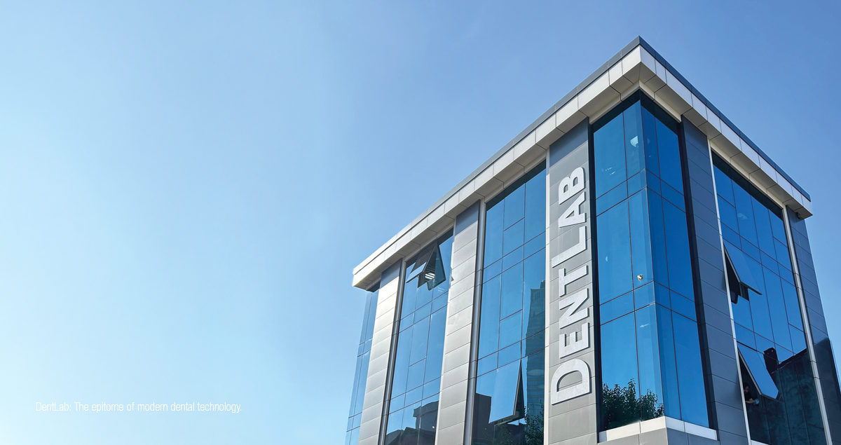
A perfect example of a laboratory with a purely digital approach is DentLab, a Turkish dental laboratory founded in 2015 by Dentgroup, Turkey’s largest Dental Service Organization (DSO). Its founder Dr Efe Celebi and his team have developed a special lab module for the group’s own practice management software, DentSoft. The module allows dental professionals to submit their orders electronically by using online forms and adding digital image data such as X-rays, intraoral scans, face scans and photographs. Users are able to see at a glance which data and information is strictly required. If desired, a user even receives procedural guidance: Specific devices like intraoral scanners, procedures and materials are recommended.


Once an order is submitted, the software sends a delivery date notification, so that the next appointment can be scheduled right away. Then, the incoming order is checked. Whenever some details are missing, the practice is contacted via a chat function in the software. Orders with incomplete or inaccurate data are rejected. A feedback function allows the technician to specify what needs to be repeated, improved or modified. Once accepted, the ordered items are produced and shipped to the office in a trackable box. It arrives with pre-treatment and cementation recommendations. Whenever necessary, patient feedback at try-in can be recorded on video or discussed live in a virtual meeting. “Digitalization has made physical distance irrelevant, and we are able to deliver high-quality restorations even if the patient is hundreds of miles away,” says Dr Efe Celebi. To facilitate improvement in the dental laboratory, the software has a case evaluation function that allows dental practitioners to evaluate the fit, function and look of every restoration.


“The most important approach to the patient is the human one. When using advanced digital technologies, we risk standardizing protocols to the extent that we forget there is an individual in front of us, with their own unique characteristics and wishes, entrusting us with their most precious asset: their smile.”
- MDT Vincenzo Castellano -


Conclusion

By establishing a well-structured, standardized and bidirectional flow of information between the dental office and laboratory, it is possible to improve the overall quality of prosthodontic treatments. It can be implemented with the aid of existing workflow management software, or set up according to individual demands using paper forms and personal meetings.


Anyway, sitting down together to discuss the topic of communication in a personal or virtual meeting has additional advantages, as it will help everyone involved develop a better understanding of the procedures carried out and challenges faced by their respective partner in the busy work environment. With this knowledge, it becomes easier to build a strong relationship that grows from mutual feedback and advice promoting common strategies for improvement. The result will be more streamlined work processes with less stress, higher-quality outcomes and happier patients.


We would like to express our gratitude to MDT Vincenzo Castellano and Dr Efe Celebi for sharing their individual approaches and the thinking behind them.


MDT Vincenzo Castellano Dr Efe Celebi


IDS 2025 – an event worth visiting!

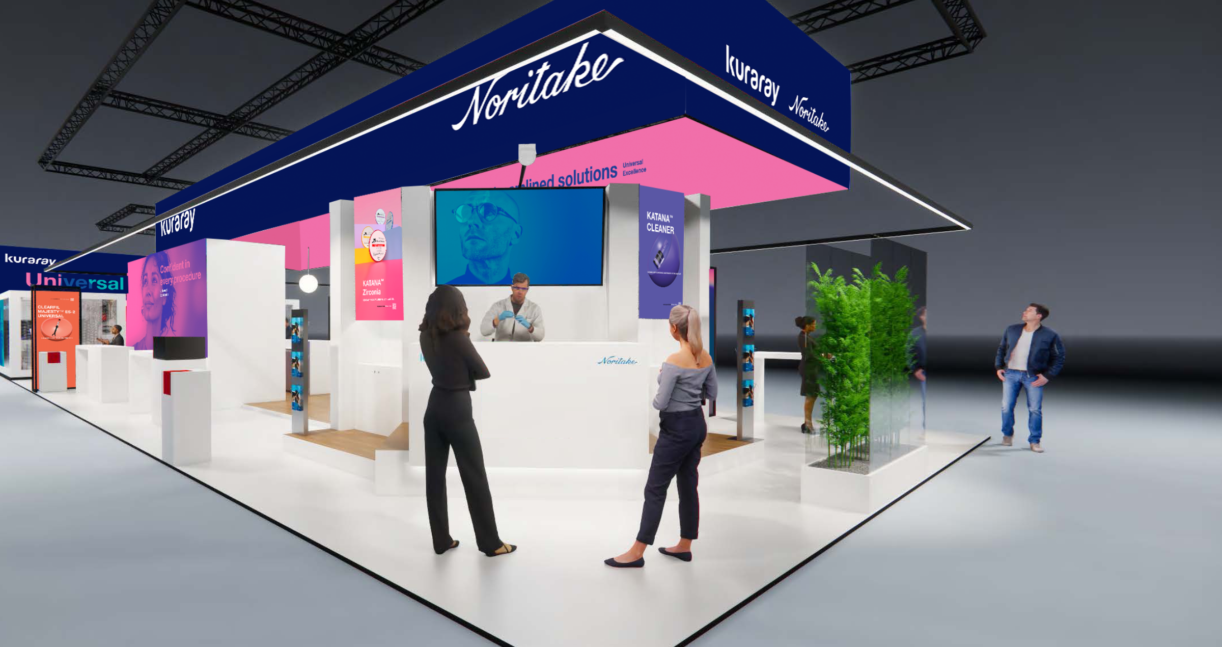
This year, the Kuraray Noritake Dental stand will be packed with news from the world of dentistry, so we offer not just one reason to visit, but a whole range: new chairside products, new labside products, inspiring lectures, enlightening hands-on demonstrations… And the best thing? You don´t have to choose just one thing, come and see them all at the Kuraray Noritake Dental booth in Hall 11.3 | Stand E010!


Chairside – smart streamlined solutions

When it comes to a dental practice, our vision is clear: a world where your materials and tools work seamlessly in your hands, where complexity is minimized, and where you are given enough time to focus on what matters most: the individual desires and needs of every single patient. This is the future of dentistry - and we are leading the way. Come discover our new additions to the UNIVERSAL EXCELLENCE family: a flowable universal composite and a new generation of a universal bond!


Labside – speed and aesthetics hand in hand

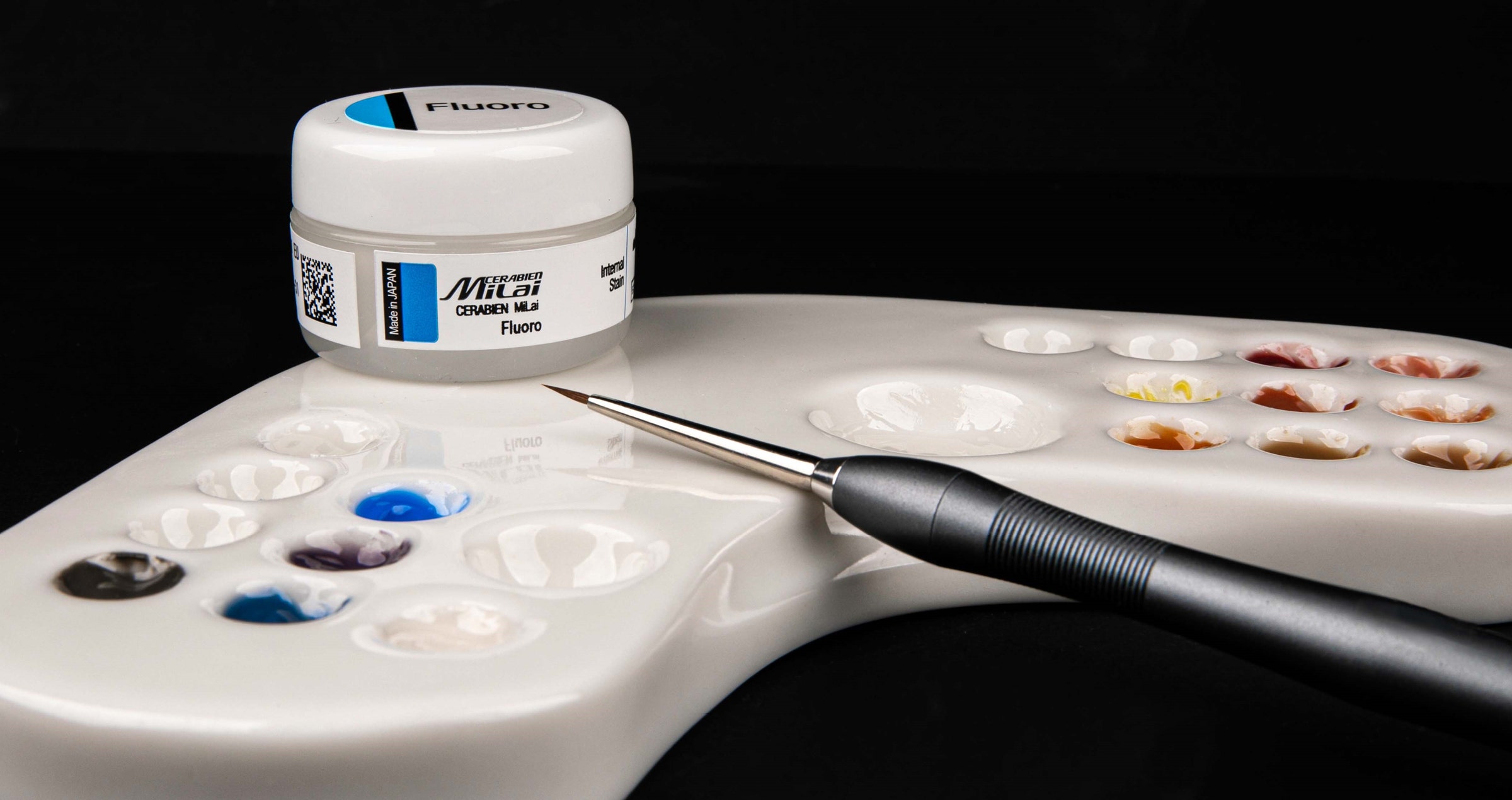
No matter whether a minimally invasive procedure or the best aesthetic outcomes are desired: Kuraray Noritake Dental has the products for you. New in the portfolio: CERABIEN™ MiLai - low-fusing porcelains and internal stains. Come and see for yourself its ultra-thin layering, exceptional mechanical properties and consistent handling for both zirconia and lithium disilicate. And while you're there, have a coffee while speed-sintering with our KATANA™ Zirconia discs takes place in real time.


Get inspired by 12 expert speakers on 2 stages

Are you a user of or interested in one of Kuraray Noritake Dental’s products or solutions, but eager to learn more about their practical use from proven experts in your field? From Wednesday until Saturday, we offer a programme of exciting lectures and live demonstrations - this time simultaneously on two stages: one dedicated to chairside and the other one to labside topics!


Warm welcome

Everyone, from sales person to scientific marketeer, looks forward to giving you a warm welcome, answering your questions and introducing you to the world of innovative Kuraray Noritake Dental products, solutions and workflows. And while you are already in Cologne anyway, why not enjoy the breathtaking beauty of the old town? Have you ever seen its famous Gothic Cathedral? Or its Old Town inviting you to stroll through its narrow alleys, finding traces of history around every corner? This year's IDS offers so many reasons to attend that it's hard not to accept the invitation.


Additional information and the full programme of lectures and hands-on courses at the IDS is available online.


Go for a trustworthy ceramic and metal primer

Article by Peter Schouten


Ceramic primers vary widely in composition and effectiveness, despite what their name might suggest. Most common primers include silane, but silane alone is insufficient for pre-treating all materials commonly used in indirect restorations before bonding.


Silane—typically in the form of γ-MPS—has a strong affinity for silica- or glass-based materials. While certain metals and their oxides can chemically react with silane, other components provide a stronger and more reliable bond to metals and metal oxides and should be seriously considered.


For metal(oxide) pretreatment in bonding, the MDP monomer is far more reactive than silane. The original MDP monomer, developed by Kuraray Co., Ltd. in 1981, remains the highest-quality MDP available, as confirmed by research.1


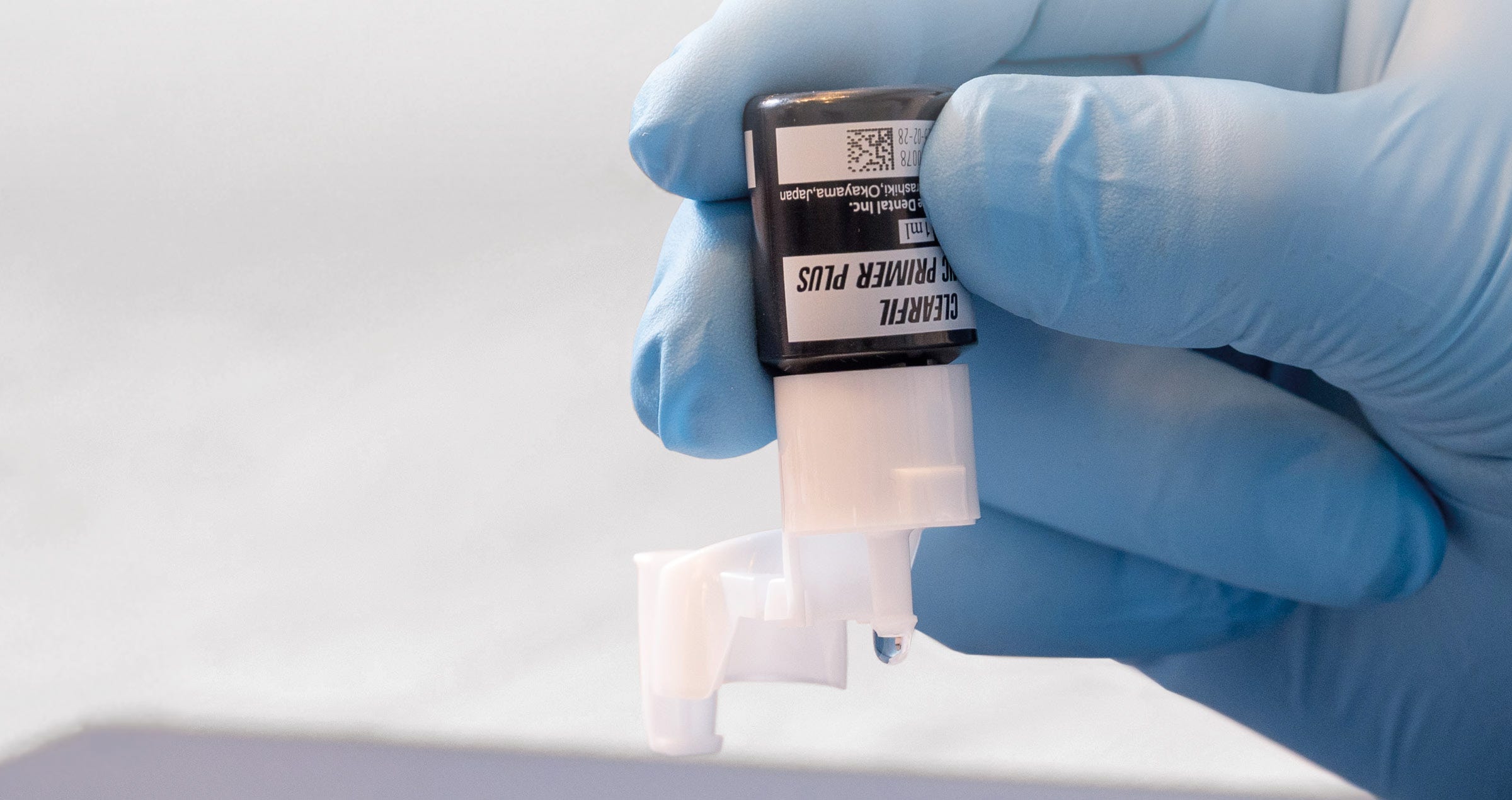
CLEARFIL™ CERAMIC PRIMER PLUS is a single-component adhesive primer that forms strong bonds with a wide range of restorative materials. This two-in-one primer incorporates the original MDP monomer, which establishes a robust bond with metals and zirconia. Simultaneously, the silane coupling agent (γ-MPS) ensures excellent adhesion to resin composites, hybrid ceramics, and glass-based ceramics such as lithium disilicate and porcelain.


ENHANCED BOTTLE DESIGN

The primer bottle is designed for effortless one-handed operation. Its unique nozzle ensures precise dispensing, minimizing the risk of contamination and spills.


PROVEN EFFECTIVENESS OF MDP-CONTAINING PRIMERS

To enhance the bonding of prosthetic materials, use a primer containing both MDP and silane. Numerous studies have demonstrated the effectiveness of this combination.


A study by Cao, Y., et al2 confirmed the superiority of CLEARFIL™ CERAMIC PRIMER PLUS (Kuraray Noritake Dental Inc.) over three other primers in improving the bond strength between zirconia and two different resin cements.

Study by Cao, Y., et al.

The effects of four primers and two cement types on the bonding strength of zirconia.

- Clearfil Ceramic Primer outperformed three other primers in improving the bond strength between zirconia and two different resin cements.

Reymus, M., et al. concluded in their study3 that before adhesive cementation of air abraded CAD/CAM resin composites, pretreatment with a specific primer, not only containing silane but also methacrylate monomers results in successfully bonded restorations. Pretreatment using an only silane containing primer results in inadequate adhesion.

Findings from Reymus, M., et al.

Bonding to new CAD/CAM resin composites: influence of air abrasion and conditioning agents as pretreatment strategy.

- Adhesive cementation of air-abraded CAD/CAM resin composites benefits from pre-treatment with a primer containing both silane and methacrylate monomers.

- Primers with only silane provided inadequate adhesion, while the combination significantly enhanced bonding success.

Four different types of CAD/CAM ceramic materials where tested in a study by Uğur, M., et al.4 Vita Mark II, IPS E.max CAD, Vita Suprinity and Vita Enamic were primed with three different primers CLEARFIL™ CERAMIC PRIMER PLUS, G-Multi Primer (GC) and Monobond S (Ivoclar Vivadent), either after hydrofluoric acid etching or no etching. It was concluded in this study that the combined effects of MDP and γ-MPTS resulted in a significant increase in the bonding strength of the resin cement to the used ceramics.

Research by Uğur, M., et al.

Effect of ceramic primers with different chemical contents on the shear bond strength of CAD/CAM ceramics with resin cement after thermal ageing.

- Four types of CAD/CAM ceramic materials (Vita Mark II, IPS e.max CAD, Vita Suprinity, and Vita Enamic) were tested with three primers: Clearfil Ceramic Primer Plus, G-Multi Primer, and Monobond S.

- Pre-treatment included hydrofluoric acid etching or no etching. Results showed that primers combining MDP and γ-MPTS substantially improved bond strength to ceramics.

Pilo, R., et al. concluded in their study5 investigating the effect of tribochemical treatment and silane reactivity on the bonding to zirconia that MDP greatly contributes to the bonding mechanism of the silane containing primers. CLEARFIL™ CERAMIC PRIMER PLUS showed to be the most reliable and effective primer in this study.

Pilo, R., et al. Study

Effect of tribochemical treatments and silane reactivity on resin bonding to zirconia.

- Examined tribochemical treatment and silane reactivity on zirconia bonding.

- MDP significantly contributed to the bonding mechanism of silane-containing primers.

- Ceramic Primer Plus was the most reliable and effective primer.

In his study6 that forms a part of his well-known dissertation, Masanao Inokoshi and others concluded that a combined mechanical/chemical pre-treatment of sintered IPS e.max ZirCAD (Ivoclar Vivadent) results in the most durable bond to zirconia. In this case when the chemical pretreatment was performed with CLEARFIL™ CERAMIC PRIMER PLUS or Monobond Plus (Ivoclar Vivadent). Scotchbond Universal (3M ESPE) and Z-PRIME Plus (Bisco), also used in this study as chemical pretreatment primers showed significant lower bond strengths.

Research by Masanao Inokoshi

Bonding effectiveness to different chemically pre-treated dental zirconia.

- Investigated mechanical/chemical pre-treatment of sintered IPS e.max ZirCAD for durable zirconia bonding.

- Clearfil Ceramic Primer (Kuraray Noritake) and Monobond Plus (Ivoclar Vivadent) yielded the most durable bonds, outperforming Scotchbond Universal (3M ESPE) and Z-PRIME Plus (Bisco), which showed significantly lower bond strengths.


OPTIMAL PRIMING FOR ADHESIVE CEMENTING

These studies underscore the critical role of MDP-containing primers in achieving reliable and durable adhesion for prosthetic materials. Products containing only silane, such as RelyX™ Ceramic Primer (3M ESPE), are less effective at creating a durable bond between resin cements or composites and ceramic- or metal-based prosthetic materials.


Image from clinical case by MDT Rondoni and Dr. Attanasio


STRAIGHTFORWARD AND EFFICIENT

The use of CLEARFIL™ CERAMIC PRIMER PLUS is straightforward: just apply it to the bonding surface, dry it, and proceed with the following treatment step. Incorporated into your process to streamline adhesion preparation and achieve reliable results!


JUST APPLY AND DRY

CLEARFIL™ CERAMIC PRIMER PLUS may be applied to any restoration surface after the required pretreatment. Pretreat the adherent surface of the restoration as indicated:


* If your laboratory already treated with a hydrofluoric acid, cleaning and activating with K-ETCHANT Syringe just before applying CLEARFIL™ CERAMIC PRIMER PLUS is recommended.

**When using with PANAVIA™ V5 or CLEARFIL™ DC CORE PLUS


Universal prosthetic primer designed for a strong bond and procedural simplicity


Author:

PETER SCHOUTEN


References

1. Yoshihara K., et al.(2015) Functional monomer impurity affects adhesive performance, Dental Materials, Volume 31, Issue 12, https://doi.org/10.1016/j.dental.2015.09.019. Pilo, R., et al. (2018). “Effect of tribochemical treatments and silane reactivity on resin bonding to zirconia.” Dent Mater 34(2): 306-316.
2. Cao, Y., et al. (2021). The effects of four primers and two cement types on the bonding strength of zirconia. Annals of Translational Medicine. 10. 10.21037/atm-21-4909.
3. Reymus, M., et al. (2019). “Bonding to new CAD/CAM resin composites: influence of air abrasion and conditioning agents as pretreatment strategy.” Clin Oral Investig 23(2): 529-538.
4. Uğur, M., et al. (2023). Effect of ceramic primers with different chemical contents on the shear bond strength of CAD/CAM ceramics with resin cement after thermal ageing. BMC Oral Health. 23. 10.1186/s12903-023-02909-z.
5. Pilo, R., et al. (2018). “Effect of tribochemical treatments and silane reactivity on resin bonding to zirconia.” Dent Mater 34(2): 306-316.
6. Inokoshi, M., et al. (2014). “Bonding effectiveness to different chemically pre-treated dental zirconia.” Clin Oral Investig 18(7): 1803-1812.


Digital workflows in dentistry and the future of dental care

Interview with Dr. Efe Celebi


In March 2024, Dr Ahmad Al-Hassiny, Director of the Institute of Digital Dentistry, shared his observations from LMT Lab Day 2024 in Chicago, noting that over 90 percent of U.S. dental laboratories and nearly 50 percent of dental practices have already adopted digital technologies and workflows. These advancements streamline the production of dental models, restorations across various materials, and much more.


Our company Kuraray Noritake Dental Inc. is dedicated not only to developing high-quality products and constantly adapting them to the needs of dentistry, but also to streamlining procedures in the dental laboratory and practice. Aiming to understand the current needs of dental technicians and dental practitioners around the world to provide what would really make a difference, we are in close dialogue with experts in the field. We love to listen to their stories, learn how digital technologies and artificial intelligence are already transforming dental procedures and see how we can contribute to a smooth transition – e.g. with products that support efficient workflows and great outcomes.


Lately, we had the chance to sit down with a leading figure in Turkey’s digital dentistry transformation, Dr Efe Celebi, to discuss the current landscape and future of digital innovation in dentistry. Being convinced that his experience is worth being shared with a broader audience, we have summarized the conversation.


Dr. Celebi, you’re known as a pioneer in digital dentistry. What drove you to establish companies with a strong digital focus?

As the founder of Dentgroup, Turkey’s largest Dental Service Organization (DSO), I have always believed in the power of digital dentistry. My goal was to establish digital workflows for producing indirect restorations, beginning with intraoral scanning at our practices. Initially, we sought laboratory partners in Turkey willing to make this transition with us. But at that time, none of our partner labs were prepared to take the leap, so we decided to build our own. Digitalization, after all, is simplifying work in almost every field, and dentistry is no exception. So, in 2015, we founded DentLab to provide cutting-edge digital laboratory services.


How did you go about implementing digital workflows?

From the very beginning, we aimed to digitize every aspect of our work—from production to data management and communication. Our practices already used our own practice management software, DentSoft, successfully. To connect our clinics with the lab, we developed a specialized lab module. This allowed our dental practitioners to submit every order electronically. It was a huge improvement over the old process, where forms with sketches were mailed physically, just as it had been in in the old days. Now, practitioners can select the teeth in need of treatment, specify restoration colour and design, and upload radiographs, photos, and intraoral scans with just a few clicks. Over time, we have continuously enhanced communication features between clinics and labs, adding things like delivery date notifications for orders, so patients can book their next appointment before they even leave the clinic. These tools have greatly improved coordination between our dentists and lab technicians.


How is communication organized in the software?

We created a chat-style communication area where different team members can talk, with all records saved and accessible to anyone involved in a treatment. This setup is a major improvement over phone calls, as every detail—from treatment notes to radiographs and photos—is stored and easy to reference. We even enabled practitioners to rate the products they receive, and every necessary remake is documented along with the responsible technician. This feedback system has allowed us to maintain high-quality standards, identify issues, and provide targeted training where needed.


Modern digital technologies used at DentLab to provide cutting-edge digital laboratory services.


Have you made other improvements in workflow and communication between clinics and laboratories?

DentLab initially served Dentgroup practices exclusively, but we eventually opened its services to other clinics. In this context, we set standards for incoming orders—from the required data to impression quality. If an order does not meet these standards, we reserve the right to reject it, explaining why, so the submitter can improve. We also implemented a loyalty programme and developed a special, trackable package with a QR code to prevent loss of items in transit. This innovation lets both the team in the lab and the clinic track each package’s location in real time, solving a common logistical challenge. We have even patented this unique packaging system.


Unique patented delivery box used at DentLab to prevent loss of items.


Do you provide guidance on materials and tools for clinics?

Yes, as part of our commitment to quality, we recommend specific intraoral scanners and even distribute them to customers outside our network. We also advise clinics on material choices, pre-treatment needs, and compatible resin cements for permanent placement of the produced restorations. Our protocols cover the entire restorative procedure. For aesthetic cases, we recommend starting with a smile design and using mock-ups. The mock-up evaluation lets practitioners and patients provide feedback, so the responsible lab technician can produce the final restoration with precision.


With digital workflows so well-established, do you think technicians still benefit from meeting patients face-to-face?

Not necessarily. Occasionally, a dentist may request a patient visit the lab, and we accommodate this. However, digitalization has made physical distance irrelevant, as clinics and labs can now work seamlessly from anywhere. In fact, we serve offices in Europe without any face-to-face interactions between patients and lab technicians. For patients, especially in a city like Istanbul, avoiding long travel times is a big plus, while we can still ensure high-quality outcomes.


Would you say digitalization has improved treatment quality overall?

Absolutely. There is a learning curve to digital processes, but once practitioners adjust, the quality is noticeably higher. In traditional workflows, practitioners might bend the rules, asking technicians to work with suboptimal impressions, for example. Digital systems do not allow for such shortcuts; preparation quality is clear from the scan, and impression errors can be corrected instantly before submitting to the lab. Additionally, digital scans will never shrink, distort, or tear during production, unlike physical impressions.


What are the main challenges associated with digital dentistry today?

The biggest challenge is simply taking the first step. Dentistry has been hesitant to change after decades of doing things the same way. Going digital requires an investment in both time and money. But those who make the switch find the rewards—better outcomes and more efficient procedures—well worth it.


What is next for digital dentistry?

With AI advancing quickly, the field is evolving in exciting ways. Today, we can combine digital impressions, facial scans, photographs, and 3D imaging to create a “virtual patient”. Some clinics are already using software to analyze digital data, like dental X-rays, and I predict that robot-assisted or even autonomous clinical procedures are on the horizon. Imagine robot arms taking impressions or patients scanning their own teeth with smartphones. Impression-taking procedures carried out at home already support aligner treatments in some cases. As these technologies advance, the need for dental assistants will likely decrease. The digital future for dentistry is incredibly promising, filled with tools that can transform patient care and practice efficiency on a global scale.


Example of beautiful, precisely fitting all-ceramic restorations produced at DentLab.


Optimizing intraoral and extraoral substrates for maximal adhesive potential

Article by Dr. Clarence Tam HBSc, DDS, FIADFE, AAACD


A NOVEL MDP-BASED SURFACTANT SOLUTION

The everyday practice of adhesive restorative dentistry, whether utilizing direct or indirect restorations, is fraught with the need for ideal environmental conditions to generate an optimal prognosis. The bonding of composite resin is the foundation of direct and indirect restoratives, as it provides the link between restoration and tooth. As dentistry strives to be minimally invasive, the treatment of the bonding interface is reflected in this philosophy by the use of self-etching multi-substrate acidic monomers such as 10-methacryloyloxyldecyl dihydrogen phosphate (10-MDP). There are myriad opportunities for both intaglio and fitting surfaces to be contaminated with varying agents to the detriment of restoration prognosis.


Some of the contaminants to be considered are of course, moisture from exhalation, ambient humidity in the oral cavity, blood, saliva and artificial sources such as provisional cement during a two-stage indirect delivery technique. Moisture is an agent which is only welcomed via a controlled approach during the dentin penetration phase of priming the substrate for adhesion, however if excessive in quantity will compromise the hybridization of the interface. Blood and saliva are ubiquitous in restorative dentistry, and best controlled via the application of rubber dam as part of an absolute isolation philosophy. Contamination of the prepared surface can also occur through artificial cements or lubrication agents. Hemostatic agents such as ferric sulfate and aluminum chloride have the ability to deposit insoluble precipitates on the surface of the tooth in a manner that 33% orthophosphoric acid can only partially remove. Also considered is the particulate deposition of dentin and enamel as part of standard tooth preparation. This smear layer is residual on the dentin surface, often occludes dentinal tubules, and is an obstacle that must be overcome in order to bond to the hydroxyapatite and collagen fibrils of the surface.


Overall, the risks to adhesive compromise and at worst, adhesive failure are high. This report details the use of a novel solution for debriding both indirect restorative and tooth intaglio with a 10-MDP salt-based solution that has the flexibility to be used both extraorally and intraorally.


ENDEMIC CONTAMINANTS: MOISTURE, BLOOD AND SALIVA

Moisture is a critical component to maximize the adhesive bond strength of certain modern universal adhesives. The presence of moisture allows for increased penetration of bonding solutions into dentinal tubules and between collagen fibrils, ultimately bolstering the resilience of the hybrid layer1. During the cementation of an indirect restoration, both salivary and blood contamination of the mating surfaces have been shown to have a deleterious effect on bond strengths, with blood contamination faring the worst in all conditions2. Van Meerbeck et al reported on technique sensitivity with one-step contemporary universal adhesives3. The basis of his findings note that these adhesives require water as an ionization medium for the self-etching reaction, with the need to evaporate water from the interfacial surfaces in order to maximize bond strengths. Despite this, these interfaces are considered semi-permeable which predisposes the hybrid layer to an increased risk of hydrolytic degradation in adhesive solutions that are not 2-hydroxyethyl methacrylate-free (HEMA-free), which has a greater affinity for water.


Periera et al tested varying degrees of wetness of dentin substrate controlled with variables such as short vs. long air blasts, wet vs. dry cotton pellets, microbrush use and an intentionally over-wet surface. In all groups, the “wettest” dentin intaglio surface resulted in the lowest shear bond strength4.


The influence of saliva and blood contamination is clearly negative in situations where the bonding interface was contaminated before or after adhesive application. For saliva, this reduction is due to the deposition of salivary glycoprotein on the surface, and relative to blood, macromolecules such as fibrinogen and platelets block access to the tubules for effective bonding. Blood contamination was found consistently to be more profoundly deleterious on bond strength relative to saliva2.


In general, on smear layer-affected dentin, chlorhexidine was consistently superior to other agents such as ethanol, EDTA, aloe vera in establishing the highest shear bond strength to dentin. On dentin that had previously been etched and contaminated with blood and saliva, the agent subsequently applied that showed the highest recovery of shear bond strength was 37.5% phosphoric acid5. A study on the nanomechanical and nanoroughness of etched dentin and self-etching adhesive treated dentin both contaminated with saliva revealed that KATANA™ Cleaner was capable of restoring control values of complex modulus and nanoroughness relative to control6.


SYNTHETIC WORKFLOW CONTAMINANTS: DENTAL STONE, HEMOSTATIC AGENTS, ROOT CANAL SEALERS AND PROVISIONAL CEMENTS

A 2020 study by Marfenko et al demonstrated that salivary contamination showed significantly lower bond strengths relative to intaglio contamination by dental stone from laboratory processes. The application of a silane coupling agent to the intaglio surface has a protective effect on the bond strength7. The caveat is that lithium disilicate-based restorations are often requested pre-etched with hydrofluoric acid from the laboratory. Often, the case is returned to the clinician on the secondary or primary model. If already treated with hydrofluoric acid, the surface can now be considered recontaminated with the stone or resin model or simply skin oils from handling. The unprotected surface needs to be decontaminated in any case following the try-in procedure, which now may feature elements of dental stone, blood and saliva, not to mention hemostatic agents such as aluminum chloride and ferric sulphate. If silane coupling agents are applied prior to try-in, the question of whether the intaglio surface was truly contaminant-free after removal from the model.


Aluminum chloride is a hemostatic agent that leaves an insoluble precipitate on the surface of the dentin, that is only partially removed when treated with phosphoric acid, resulting only in a partial recovery of shear bond strength relative to control. The application of ethylene diacetyl tetrasodium acetate (EDTA) returned the bond strengths to the level of normal dentin8. The bonding of polycrystalline ceramic restorations and metal alloys is contaminated with saliva upon try-in. This can be removed via steam cleaning and air particle abrasion set at 2.5 bar for 15 seconds9. Phosphoric acid is often mistakenly applied as a cleaning agent to the intaglio surface. In polycrystalline ceramics such as tetragonal zirconia polycrystal, this is disastrous, as phosphates will bond firmly to the free sites that the 10-MDP monomer normally bonds to as part of the APC protocol of zirconia bonding, significantly compromising bond strength (Blatz, 2016)10. A study of modern surface cleaners demonstrated successful debridement of the surface using KATANA™ Cleaner for both blood and saliva-contaminated substrates11, 12.


Provisional cements are thought to have a deleterious effect on the shear bond strength of adhesively-bonded indirect ceramic restorations. Ding et al (2022) uncovered that resin-based and non-eugenol cement use in the provisional phase decreased the bond strength relative to control, whilst the use of calcium hydroxide and polycarboxylate cements exhibited acceptable metrics. Debridement of the prepared surface with air particle abrasion (APA) resulted in recovery of decreased bond values to that of control13. Equally useful was the application of Immediate Dentin Sealing (IDS)14, a technique characterized ideally by APA before adhesive bonding and the application of a resin coat, occluding both the dentin tubules as well as the oxygen inhibition layer, allowing the resin-dentin bond to mature and strengthen in the absence of stresses. This approach is effective in minimizing post-operative hypersensitivity and bacterial ingress, as well as optimizing the shear bond strength particularly when indirect ceramics are concerned15. Hardan et al found that the shear bond strength was highest when IDS was completed using a three-step etch and rinse adhesive protocol14.


Hemostatic agents used in clinical dentistry exhibit a pH of 1.1 to 3.0 and are as acidic as self-etching primers16. Chaibutyr and Kois found that dentin when contaminated with 25% aluminum chloride or 13% ferric sulphate demonstrated a significantly lower shear bond strength to dentin, which was significantly recovered using the etch-and-rinse approach17. This approach albeit successful was only able to achieve partial reversal of shear bond strength deficits relative to control, with a pre-etching application of EDTA required in order for full recovery8. KATANA™ Cleaner was found to have a positive effect on the cleaning of dentin contaminated with both aluminum chloride and ferric sulphate.


The bonding of dentin substrate contaminated with root canal sealers is a concern for the integrity of core buildups post-endodontic treatment. The use of  KATANA™ Cleaner was found to be generally superior to the ethanol test subgroup in the removal of zinc-oxide eugenol-based sealer with equal performance to 70% ethanol for the epoxy resin-based sealer18.


CLINICAL CASE DEMONSTRATION

A 35 year old ASA 1 female patient presented to the practice with multiple failing composite restorations in the second quadrant that were planned for replacement. Prior to the delivery of topical and local anaesthesia, it is common procedure in the practice to ascertain shade specifics of planned restoratives before potential dehydration can affect the optical properties of the natural tooth. Smart monochromatic composites (Fig. 1) are a class of direct restoratives that leverages the ability of its nanofiller composition and refractive index to mimic the structural color of the surrounding enamel and dentin19. This typically enables a clinician to have a simplified selection of shades on hand.


Two carpules of 2% Lignocaine with 1:100,000 epinephrine were delivered via buccal infiltration before absolute isolation was achieved using a non-latex rubber dam (Isodam HD Heavy, 4D Rubber, UK) (Fig. 2). The old restorations were excavated along with caries (Fig. 3), and the dentin structure assessed for residual decay with a detector dye (Caries Detector, Kuraray Noritake Dental Inc.). The preparation cavosurface margins were gently bevelled before surface treatment with air particle abrasion (30psi, 29 micron aluminum oxide in a 17.5% ethanol carrier, Aquacare UK) (Fig. 4). The enamel margins were etched with 33% orthophosphoric acid and rinsed (Fig. 5). The preparation surfaces were decontaminated further of any residual smear or powder residue using a MDP-based surfactant (KATANA™ Cleaner, Kuraray Noritake Dental Inc.) (Fig. 6). A single step self-etching universal adhesive was applied to the preparation as per manufacturer instructions and air thinned before light curing (Fig. 7).


Fig. 1


Fig. 2


Fig. 3


Fig. 4


Fig. 5


Fig. 6


Fig. 7


A matrix-in-matrix approach was utilized for the second bicuspid as the first step to allow for simultaneous anatomic construction of the mesial and distal marginal ridges. This technique does not require the use of a wedge as the outer circumferential Tofflemire matrix (Omnimatrix, Ultradent Products) tightens cervically around the inner anatomically-curved sectional matrix (Garrison Firm Band, Garrison Dental Solutions) allowing for a hermetic gingival seal (Fig. 8). If required, the setup may be further modified by the use of Teflon inserted between the two matrices to provide better proximofaciolingual adaptation. As a result, finishing and contour creation post-band removal is kept to a minimum. Following this, a traditional sectional matrix system may be employed to close contacts and build marginal ridges in the conventional manner (Fig. 9).


Following marginal ridge construction, the matrix assembly was removed and with the Class II lesions converted into a Class I situation, microlayering proceeded with a high flexural strength flowable liner (CLEARFIL MAJESTY™ Flow, Kuraray Noritake Dental Inc.) prior to the application of a monochromatic composite resin (CLEARFIL MAJESTY™ ES-2 Universal U shade, Kuraray Noritake Dental Inc.). The buccal cusps were constructed first as the author considers this essential to establishing restoration lobe proportions (Fig. 10). Subsequent layers were completed in a lobe-by-lobe approach to finish the occlusal anatomy (Fig. 11 and 12). The restoration was checked for occlusal functional conformativity, finished and polished to high shine (Fig. 13).


Fig. 8


Fig. 9


Fig. 10


Fig. 11


Fig. 12


Fig. 13


DISCUSSION

Dental substrates are often contaminated in both direct and indirect restorative processes. Historically, etch-and-rinse approaches have been successful for at least the partial recovery of bond strength however it is not practical in situations where selective or self-etching is the adhesive strategy. The restorative dentist in these cases can use the 10-MDP monomer in self-etching systems to target non-demineralized dentin such as CLEARFIL™ Universal Bond Quick to establish an acid base resistance zone (ABRZ) otherwise known as Super Dentin20. The presence of the operative smear layer impedes full access of the self-etching primer to the dentin substrate in some cases. In such cases, without KATANA™ Cleaner, APA is required to transform the substrate back to control bonding potential. APA as a modality is only utilized by a subset of dental practitioners often due to financial constraints or lack of technique experience. KATANA™ Cleaner thus represents a versatile tool for the decontamination and  optimization of substrate surfaces for adhesive bonding both in intraoral and extraoral applications. Its ability to re-establish ideal bonding values in areas that are not effectively reached by APA such as endodontic canal anatomy in a non-invasive manner literally cements it as a truly indispensable tool for the modern restorative dentist.


Disclaimer: Some indications are not described in the product’s Instructions for Use and are based on published research and/or the author’s experience. Before using each product, read carefully the Instructions for Use supplied with the product for full details and workflows.


Dentist:

CLARENCE TAM


References

1. Sugimura R, Tsujimoto A, Hosoya Y, Fischer NG, Barkmeier WW, Takamizawa T, Latta MA, Miyazaki M. Surface moisture influence on etch-and-rinse universal adhesive bonding. Am J Dent. 2019 Feb;32(1):33-38. PMID: 30834729.
2. Taneja S, Kumari M, Bansal S. Effect of saliva and blood contamination on the shear bond strength of fifth-, seventh-, and eighth-generation bonding agents: An in vitro study. J Conserv Dent. 2017 May-Jun;20(3):157-160. doi: 10.4103/0972-0707.218310. PMID: 29279617; PMCID: PMC5706314.
3. Van Meerbeek B, Van Landuyt K, De Munck J, Hashimoto M, Peumans M, Lambrechts P, Yoshida Y, Inoue S, Suzuki K. Technique-sensitivity of contemporary adhesives. Dent Mater J. 2005 Mar;24(1):1-13. doi: 10.4012/dmj.24.1. PMID: 15881200.
4. Pereira GD, Paulillo LA, De Goes MF, Dias CT. How wet should dentin be? Comparison of methods to remove excess water during moist bonding. J Adhes Dent. 2001 Fall;3(3):257-64. PMID: 11803713.
5. Haralur SB, Alharthi SM, Abohasel SA, Alqahtani KM. Effect of Decontamination Treatments on Micro-Shear Bond Strength between Blood-Saliva-Contaminated Post-Etched Dentin Substrate and Composite Resin. Healthcare (Basel). 2019 Nov 1;7(4):128. doi: 10.3390/healthcare7040128. PMID: 31683858; PMCID: PMC6956069.
6. Toledano M, Osorio E, Espigares J, González-Fernández JF, Osorio R. Effects of an MDP-based surface cleaner on dentin structure, morphology and nanomechanical properties. J Dent. 2023 Nov;138:104734. doi: 10.1016/ j.jdent.2023.104734. Epub 2023 Oct 2. PMID: 37793561.
7. Marfenko S, Özcan M, Attin T, Tauböck TT. Treatment of surface contamination of lithium disilicate ceramic before adhesive luting. Am J Dent. 2020 Feb;33(1):33-38. PMID: 32056413.
8. Ajami AA, Kahnamoii MA, Kimyai S, Oskoee SS, Pournaghi-Azar F, Bahari M, Firouzmandi M. Effect of three different contamination removal methods on bond strength of a self-etching adhesive to dentin contaminated with an aluminum chloride hemostatic agent. J Contemp Dent Pract. 2013 Jan 1;14(1):26-33. doi: 10.5005/jp-journals-10024-1264. PMID: 23579888.
9. Yang B, Lange-Jansen HC, Scharnberg M, Wolfart S, Ludwig K, Adelung R, Kern M. Influence of saliva contamination on zirconia ceramic bonding. Dent Mater. 2008 Apr;24(4):508-13. doi: 10.1016/j.dental.2007.04.013. Epub 2007 Aug 6. PMID: 17675146.
10. Blatz MB, Alvarez M, Sawyer K, Brindis M. How to Bond Zirconia: The APC Concept. Compend Contin Educ Dent. 2016 Oct;37(9):611-617; quiz 618. PMID: 27700128. (7)
11. Awad MM, Alhalabi F, Alzahrani KM, Almutiri M, Alqanawi F, Albdiri L, Alshehri A, Alrahlah A, Ahmed MH. 10-Methacryloyloxydecyl Dihydrogen Phosphate (10-MDP)-Containing Cleaner Improves Bond Strength to Contaminated Monolithic Zirconia: An In-Vitro Study. Materials (Basel). 2022 Jan 28;15(3):1023. doi: 10.3390/ma15031023. PMID: 35160968; PMCID: PMC8838745.
12. Tian F, Londono J, Villalobos V, Pan Y, Ho HX, Eshera R, Sidow SJ, Bergeron BE, Wang X, Tay FR. Effectiveness of different cleaning measures on the bonding of resin cement to saliva-contaminated or blood-contaminated zirconia. J Dent. 2022 May;120:104084. doi: 10.1016/j.jdent.2022.104084. Epub 2022 Mar 3. PMID: 35248674.
13. Ding J, Jin Y, Feng S, Chen H, Hou Y, Zhu S. Effect of temporary cements and their removal methods on the bond strength of indirect restoration: a systematic review and meta-analysis. Clin Oral Investig. 2023 Jan;27(1):1530. doi: 10.1007/s00784-022-04790-6. Epub 2022 Nov 24. PMID: 36422719; PMCID: PMC9877054.
14. Hardan L, Devoto W, Bourgi R, Cuevas-Suárez CE, Lukomska-Szymanska M, Fernández-Barrera MÁ, Cornejo Ríos E, Monteiro P, Zarow M, Jakubowicz N, Mancino D, Haikel Y, Kharouf N. Immediate Dentin Sealing for Adhesive Cementation of Indirect Restorations: A Systematic Review and Meta-Analysis. Gels. 2022 Mar 11;8(3):175. doi: 10.3390/gels8030175. PMID: 35323288; PMCID: PMC8955250.
15. Samartzi TK, Papalexopoulos D, Sarafianou A, Kourtis S. Immediate Dentin Sealing: A Literature Review. Clin Cosmet Investig Dent. 2021 Jun 21;13:233-256. doi: 10.2147/CCIDE.S307939. PMID: 34188553; PMCID: PMC8232880.
16. Woody RD, Miller A, Staffanou RS. Review of the pH of hemostatic agents used in tissue displacement. J Prosthet Dent. 1993 Aug;70(2):191-2. doi: 10.1016/0022-3913(93)90018-j. PMID: 8371184.
17. Chaiyabutr Y, Kois JC. The effect of tooth-preparation cleansing protocol on the bond strength of self-adhesive resin cement to dentin contaminated with a hemostatic agent. Oper Dent. 2011 Jan-Feb;36(1):18-26. doi: 10.2341/09-308-LR1. Epub 2011 Feb 21. PMID: 21488725.
18. Tian F, Jett K, Flaugher R, Arora S, Bergeron B, Shen Y, Tay F. Effects of dentine surface cleaning on bonding of a self-etch adhesive to root canal sealer-contaminated dentine. J Dent. 2021 Sep;112:103766. doi: 10.1016/j.jdent.2021.103766. Epub 2021 Aug 5. PMID: 34363888.
19. Ahmed MA, Jouhar R, Khurshid Z. Smart Monochromatic Composite: A Literature Review. Int J Dent. 2022 Nov 8;2022:2445394. doi: 10.1155/2022/2445394. PMID: 36398065; PMCID: PMC9666026.
20. Nikaido T, Weerasinghe DD, Waidyasekera K, Inoue G, Foxton RM, Tagami J. Assessment of the nanostructure of acid-base resistant zone by the application of all-in-one adhesive systems: Super dentin formation. Biomed Mater Eng. 2009;19(2-3):163-71. doi: 10.3233/BME-2009-0576. PMID: 19581710.


Simplifying esthetic composite reconstructions using CLEARFIL MAJESTY™ ES-2 Universal

Article by Dr. Clarence Tam HBSc, DDS, FIADFE, AAACD


A CHAMELEON SUPERCOMPOSITE


INTRODUCTION

The name of the game in modern-day esthetic and restorative dentistry is that of Responsible Esthetics. The goal of treatment typically strives to correct any structural and cosmetic shortfalls in both biologically-driven and trauma-affected teeth with the precise, artistic placement of various replacement layers, all whilst respecting and retaining a maximal volume of residual tooth structure. Anterior teeth can be affected by enamel and dentin dysplasia, caries and sclerotic conditions and are characterized by a laundry list of genetically-derived and environmentally-acquired conditions with an esthetic deficit that often threaten an individual’s functional and psychosocial integrity if not restored to the seamless picture of health.


Missing and defective tooth structure must be categorized into its attendant enamel and dentin components. Both substrates are distinctly different in composition, with enamel being highly inorganic in nature and dentin proportionately more collagenous in nature. The latter stratum is responsible for the refraction of light, the expression of the true color of the tooth, namely the hue and the endowment of fracture toughness or resilience in functional performance. The value and chroma are the other elements of color and are modified by the thickness of enamel. The replacement of enamel has been found to be best substituted from a biomechanical perspective by adhesively-bonded indirect porcelain restorations, and dentin using both composite resin and short fiber reinforced composite (SFRC), the latter imparting increased fracture toughness in large volume replacement restorations, especially those with pericervical structural deficits.


In adolescent patients, the gold standard of treatment involves direct composite resin, as often zero to minimal tooth structure preparation is required as a foundation to the bonded restorative. It would be impractical to use bonded indirect restorations when the development of the dentition in puberty is continuous, especially with the retraction of gingiva as one progresses to young adulthood. Resin composite allows prescience in the opportunity to predictably modify and/or add to the existing restoration if dental bleaching for the other teeth is desired or if a further traumatic incident is encountered. The ability to modify bonded porcelain is not predictable and frequent marginal failures occur due to a lower shear bond strength to bonded composite, especially after thermocycling. This is despite our ability to establish a chemical linkage via silane coupling agents from silicate ceramics to resin composite especially at a blended interface.


STATEMENT OF PROBLEM

Dental shades in clinical dentistry have long been classified using the VITA* Classical A1 – D4 shade guide. Despite being ubiquitous in dental practices, composite resin systems with corresponding shade systems do not satisfactorily match to their purported shade1. Floriani et al found that various mixtures of different shades in one system was required to achieve an acceptable color match with the VITA* Classical shades using the CIEDE2000 formula. Testing another composite resin, they found that none of the A1, A2 or A3 shades matched acceptably to the standard shade guide2. Indeed, even with indirect ceramic layering systems, a wide range of unacceptable discrepancy was noted between VITA* labeled porcelain shades and the actual shade guide3. The VITA* Classical shade guide became the standard in dental shade classification with the release of its A1-D4 shade guide in 1985. The majority of human-tested dental shades has been found to be in the A-family (78.5%), followed by C (13.2%), D (5.2%) and B (3.1%)1. As such, the shade accuracy of a given composite system must be important if they are to be visually naturomimetic.


CHAMELEON EFFECT DEVELOPMENT

There are myriad composite resin systems featuring a simplified shade Universal system that have acceptable chameleon effects due to their balance of translucency, light transmission, diffusion and refractive index properties. There is a concern over how these optical properties may change after both thermocycling and wet storage, potentially compromising the excellent initial esthetic blend4. Refractive index (RI) is best optimized when the RI of the inorganic fillers match closely with the RI of the cured organic matrix, typically in a range between 1.47 and 1.525. If the match is dissimilar, this drives up the opacity of the restoration due to heightened refraction and reflection at the filler/matrix interface6.


Layering of composite to mask an intraoral defect is complicated by the need to mask any linear defects such as fracture lines superimposed over the shadowing of the dark intraoral cavity in addition to regional color variations. It is confounded by the requirement to recreate natural maverick and translucent effects particularly in the incisal window region of upper and lower incisors and canines, giving the illusion of a virgin, healthy tooth. This has been historically difficult to accomplish in anterior teeth given the need to block out restorative interfaces with natural tooth structure and recreate a seamless internal structure and details. This detailed layer belies a well contoured enamel layer with realistic translucency, polishability and accurate primary and secondary anatomy.


Adding to the complexities described above, the histoanatomical approach to composite layering dictates that missing enamel is replaced by enamel shades, and dentin by the corresponding dentin shade in the appropriate shade. This shade must be selected at the very start of the appointment, as often even a minute of dehydration has a negative effect on both the perceptibility threshold and acceptability threshold of teeth7, resulting in the incorrect shade.


DEVELOPMENT

CLEARFIL MAJESTY™ ES-2 is a value-based super-nanofilled composite system that covers 15 VITA* shades in just 4 shade options with its Universal series. This Universal series provides a chameleon effect and has 4 variants: Universal (U), Universal Light (UL), Universal Dark (UD) and Universal White (UW). It is the VITA*-approved shading concept relative to color accuracy. Incorporating nano-fillers that consist of silanated barium glass fillers and slanted silica nanoclusters, its wear resistance is high and features minimal abrasiveness against the functional antagonist. The RI of both inorganic filler and organic matrix are well-matched, and the high refractive index of the composite mimics and is extremely similar to natural enamel (1.613) and dentin (1.540), thanks to an innovation labeled Light Diffusion Technology (LDT), which distorts light in a similar way dental tissue does8. There is comfort that the stability of refractive index and other optical transmission properties remains statistically stable even after artificial thermocycling and water-storage aging studies4. The color stability of CLEARFIL MAJESTY™ ES-2 has been proven over time, where a direct comparison to Filtek Ultimate showed CLEARFIL MAJESTY™ ES-2 to feature significantly less color variation from baseline and marginal functional wear over a three to four year period in teeth featuring amelogenesis imperfecta9. This color substantivity is important as dietary and environmental stressors applied over time should have as minimal effect on the restoration to ensure continued esthetic integration.


CLINICAL PROTOCOL

CLEARFIL MAJESTY™ ES-2 Universal is a monochromatic solution that covers the five key shades featured in the CLEARFIL MAJESTY™ ES-2 Premium. As such, it exhibits the most significant LDT relative to all five shades, as its ability is equal when blending to higher value translucent shades as it does to cervical chromatic shades. In a Class IV restoration with a defined fracture line, the challenge is to restore the tooth in a minimal volume of available space. The alchemy requires a complete visual occlusion of the fracture line position, and recreation of internal and external opaque and translucent anatomy along with maverick staining, craze lines and effects. In anterior teeth, the idiom of “the less you see, the less you notice” is not true, especially due to the presence of incisal edge window effects as above, however, materials with the best light diffusion and structure transference properties should be utilized to ensure the highest probability of success.


A 15 year old ASA I female presented to the practice exhibiting aged, chromatic composite restorations with poor marginal integration and gross axial overhangs; essentially a gross failure of primary anatomy and esthetics. She had been involved in a bike accident where she high-sided off braking sharply in a face-meets-concrete scenario, resulting in an uncomplicated moderate enamel-dentin fracture with blushing, affecting both the facial and palatal aspects of tooth 1.1 and a mild uncomplicated enamel dentin fracture affecting the distoincisobuccolingual aspect of tooth 2.1. The restoration overhangs were significant, extending into the proximal contour zone, thus obviating effective interdental cleaning. Vitality tests were confirmed along with radiographs to exclude the presence of apical pathology. The patient accepted the option of pre-prosthetic whitening, to improve the value characteristics of the adjacent teeth, allowing the selection of a brighter value shade combination. Intraoral digital scans were acquired and custom bleaching trays with a no reservoir, cervical seal-priority design were fabricated. The patient was instructed to bleach overnight for a 2 week period using a 10% carbamide peroxide solution (Opalesence, Ultradent Products, UT) until her maximal value was reached. Her baseline shade of the incisors was a 1M1/2M1 combination in the upper incisors and a 2M1 in the lower incisors. On final post-bleach assessment she exhibited a lightened shade of VITA* 0M3 in all incisors. The patient was instructed to use a fluoride-containing, amorphous calcium phosphate complex (ToothMousse Plus, GC America) during the following 2 weeks after cessation of whitening whilst the residual oxygen radical species dissipated from the teeth.


Fig. 1. Pre-operative unrestricted smile 1:2 ratio view, teeth 1.1 and 2.1 with old, defective composite restorations with excessive chroma.


On the day of the procedure, the pre-dehydrated shade was assessed using the supplied “real composite” shade guide tabs featured in the CLEARFIL MAJESTY™ ES-2 Premium system, with the enamel shade being WE (White Enamel) and the dentin shade WD (White Dentin). It was assessed that both white maverick effects as well as a moderate halo effect was desired along with moderate to strong translucency in the incisal window.


The patient was anesthetized using 1.5 carpules of 2% Lignocaine with 1:100,000 epinephrine (Septodont) before a rubber affixed with individual ties for the central incisors (NicTone Medium). Excavation of the old restorative material was undertaken, and the residual natural incisal edge was found to be undermined by a through-and-through fracture. Thus, the preparation was converted into a true Class IV design, with the facioincisal cavosurface margin subjected to an infinity bevel. The maxillary central incisors were isolated from the lateral incisors by way of a serrated metal strip (Komet) and the prepared surfaces subjected to micro particle abrasion using a 29 micron aluminum oxide powder in 17.5% ethanol carrier (Aquacare). The surfaces were subsequently treated with a calcium sodium phosphosilicate powder (Sylc, Aquacare) to increase the inorganic content of the prepared surface especially extending into the exposed tubules. The teeth were etched using a 33% orthophosphoric acid before a 1 minute 2% chlorhexidine scrub (Vista Products). The surface was reduced to a moist dentin surface before the bond applied, air thinned and cured.


A Mylar strip was pre-crimped in the palatoproximal line angles and positioned on the linguoaxial surface of both teeth 1.1 and 2.1. There is no shade guide for the CLEARFIL MAJESTY™ ES-2 Universal U shade, as it bears a significant chameleon effect however it does come in a light (L) and dark (D) variant. The UL shade was deemed the most suitable for the palatal or lingual shelf, with an average thickness of 0.3mm. This layer was applied in a freehand fashion with a focus on establishing the desired outline form of the tooth relative to the contralateral 2.1. The Mylar matrix setup was removed and a precurved metal matrix (Garrison Slickband, Garrison Dental) was oriented in a position perpendicular to its normal placement interproximally, and the end of the curved band tucked into the sulcus before being secured by a wedge. In this way, there is light separation of the central incisors and an intimate contact between the matrix band and the mesial edge of the freshly applied lingual shelf. A 0.5mm frame extending more than halfway through the contact point was created and cured. The process was repeated on tooth 2.1 with the goal of recreating both lingual and proximal walls of the restoration, leaving only the facial volume to be replaced.


Fig. 2. Pre-crimped Mylar matrix repeated on the DIBP aspect of tooth 2.1 to close the available space. CLEARFIL MAJESTY™ ES-2 Universal UL is used here.


Block-out of the composite extensions against the natural tooth structure was achieved by opacification using an opaque composite resin (WD, CLEARFIL MAJESTY™ ES-2 Premium, Kuraray Noritake Dental Inc.) layered in both horizontal and vertical increments. It is noted that the restorative join line must be completely obscured at the end of layering the dentin volume, otherwise the case will have almost certain esthetic failure. The internal dentin anatomy and its inherent variation was created to mirror that of the 2.1, which had minimal compromise of its incisal window with details intact. A super translucent composite resin (Clear, CLEARFIL MAJESTY™ ES-2 Premium, Kuraray Noritake Dental Inc.) was placed between the lobes of the dentin layers and cured. A 9:1 ratio of white: orange tint was mixed and placed on the incisal edge and proximoincisal corners to recreate the halo effect. A pure white tint was placed in gentle dentin mamelon-connecting spider legs up to the incisal edge to impart the realism. This was layered in a manner consistent with the appearance of the 2.1.


Fig. 3. Both horizontal and vertical dentin composite increments are demonstrated mimicking the contralateral tooth.


Fig. 4 & 5. Final immediate post-operative result after finishing and polishing.


DISCUSSION

The esthetic merit of this case is foundationally supported by composite resin technology on multiple levels. The color and physical stability over time needs to be proven in order for the clinician to have faith in its prognostication. Specifically, the material needs to have an excellent and well-matched refractive index, and one that is unaffected by both water and thermocycling stressors.


The palatal shelf was fabricated using a new-generation super nano-filled universal composite system that boasts a strong chameleon effect. If it is our intention to fool the eye, to obscure, then this first layer works well to start the blockout process of the darkness of the mouth behind the fracture line of the restored tooth. Following this, the chroma and value of the tooth are corrected using the dentin, simultaneous to its continued opacification of the fracture line and intraoral darkness. Both dentin and enamel layers are applied histoanatomically, that is, in a manner respecting the various thickness zones observed in nature.


Ultimately, esthetic success in direct composite resin is not dictated on the first day post-operatively. Factors are in play, from dehydration to occlusal wrinkles that need to be ironed out and corrected. The win depends on what material is used, along with how that material was developed to what standards, and why shade accuracy is so important in a world of variety. In a dental world with myriad composite options, we are looking for precision. Precision in technology leads to efficiency and physicoesthetic maintenance in clinical results. This ultimately results in a boost to clinician-patient confidence and an optimal prognosis.

Dentist:

CLARENCE TAM


*VITA is a trademark of VITA Zahnfabrik, Bad Sackingen, Germany


References


1. Elamin HO, Abubakr NH, Ibrahim YE. Identifying the tooth shade in group of patients using Vita Easyshade. Eur J Dent. 2015 Apr-Jun;9(2):213-217. doi: 10.4103/1305-7456.156828. PMID: 26038652; PMCID: PMC4439848.
2. Floriani F, Brandfon BA, Sawczuk NJ, Lopes GC, Rocha MG, Oliveira D. Color difference between the vita classical shade guide and composite veneers using the dual-layer technique. J Clin Exp Dent. 2022 Aug 1;14(8):e615-e620. doi: 10.4317/jced.59759. PMID: 36046166; PMCID: PMC9422970.
3. Gurrea J, Gurrea M, Bruguera A, Sampaio CS, Janal M, Bonfante E, Coelho PG, Hirata R. Evaluation of Dental Shade Guide Variability Using Cross-Polarized Photography. Int J Periodontics Restorative Dent. 2016 Sep-Oct;36(5):e76-81. doi: 10.11607/prd.2700. PMID: 27560681.
4. Almasabi W, Tichy A, Abdou A, Hosaka K, Nakajima M, Tagami J. Effect of water storage and thermocycling on light transmission properties, translucency and refractive index of nanofilled flowable composites. Dent Mater J. 2021 May 29;40(3):599-605. doi: 10.4012/dmj.2020-154. Epub 2020 Dec 24. PMID: 33361663.
5. Arai Y, Kurokawa H, Takamizawa T, et al.. Evaluation of structural coloration of experimental flowable resin composites. J Esthet Restor Dent. 2020;e12674.
6. Ota M, Ando S, Endo H, et al.. Influence of refractive index on optical parameters of experimental resin composites. Acta Odontol Scand. 2012;70(5):362–367.
7. Suliman S, Sulaiman TA, Olafsson VG, Delgado AJ, Donovan TE, Heymann HO. Effect of time on tooth dehydration and rehydration. J Esthet Restor Dent. 2019 Mar;31(2):118-123. doi: 10.1111/jerd.12461. Epub 2019 Feb 23. PMID: 30801926.
8. Meng Z, Yao XS, Yao H, Liang Y, Liu T, Li Y, Wang G, Lan S. Measurement of the refractive index of human teeth by optical coherence tomography. J Biomed Opt. 2009 May-Jun;14(3):034010. doi: 10.1117/1.3130322. PMID: 19566303.
9. Tekçe N, Demirci M, Sancak EI, Güder G, Tuncer S, Baydemir C. Clinical Performance of Direct Posterior Composite Restorations in Patients with Amelogenesis Imperfecta. Oper Dent. 2022 Nov 1;47(6):620-629. doi: 10.2341/21-106-C. PMID: 36281978.


Wishing you a wonderful Holiday Season!

2025 MARKS THE YEAR OF THE SNAKE

We wish you a successful New Year and hope you will join us on our journey in 2025.


WISHING YOU AN INNOVATIVE YEAR!


Individualisation of monolithic zirconia restorations

Article by Dr. Florian Zwiener


Modern multi-layered zirconia such as KATANA™ Zirconia STML (Kuraray Noritake Dental Inc.) already meets high aesthetic demands due to its natural colour gradient and high translucency. To achieve further characterisation and optical adjustment to the adjacent teeth, there are essentially two options: veneering with feldspathic ceramic or glazing and individualisation with ceramic stains.


While there are still many indications for veneering, especially in the anterior area, more and more cases can now be solved with monolithic restorations. This allows for a time-efficient chairside workflow with same-day treatment, eliminating the need for temporary restorations. Additionally, the absence of a porcelain layer reduces the wall thickness of the restoration and thus the space required, allowing for less invasive preparation. This also reduces the risk of endodontic complications induced by tooth preparation (grinding trauma). Another advantage is a significant reduction in the chipping risk.


Below are the essential steps for individualisation using ceramic stains, demonstrated through the example of a molar crown.


PREPARATION

The restoration is designed in full contour as usual, ideally dry-milled, and then sintered. After sintering, the restoration is first sandblasted (aluminium oxide 50 μm, 1 to 1.5 bar pressure). This microscopic roughening of the ceramic surface enables an optimal bond with the glaze. Subsequently, the restoration should be cleaned using a steam cleaner or an ultrasonic cleaner to remove all blasting residue.


The functional restoration surfaces must then be polished to avoid the risk of excessive abrasion on the enamel of the opposing dentition, as zirconia is harder than enamel. Following this, optional glazing and characterization with ceramic stains can be performed. However, for areas not in the aesthetic zone, such as the palatal surfaces of maxillary anterior teeth, this is not necessarily required.


PREPARATION: STEPS AT A GLANCE

  1. Sandblasting of the sintered restoration (Al2O3 50 μm, 1-1.5 bar)
  2. Cleaning (steam cleaner or ultrasonic cleaner)
  3. Polishing the occlusal/palatal contact areas


Fig. 1. Sintered and sandblasted zirconia crown.


Fig. 2. Occlusal high-gloss polish.


Fig. 3. TWIST™ DIA for Zirconia (Kuraray Noritake Dental Inc.) enables efficient polishing of zirconia in three steps.


STAINING AND GLAZING

The shades A+, B+, C+, and D+ of the paste-like ceramic stain CERABIEN™ ZR FC Paste Stain (Kuraray Noritake Dental Inc.) enhance the chroma in the cervical area when applied in the respective tooth shade. They are used to strengthen the multicolour effect of the zirconia or to darken the restoration overall. By mixing the stains with glaze or clear glaze in different ratios, the intensity can be adjusted.


Cervical 1 and 2 are suitable for replicating exposed cervical areas or discolouration. Cervical 1 is also useful for marking fissures, as it gives the crown depth and structure without appearing overly dark. Patients typically reject excessively pronounced fissure effects. Since fissure areas in multi-layered materials generally lie in the lightest part of the block (in the enamel layer), it may make sense to darken them slightly with A+, while white hypermineralisations can be replicated on the cusp tips. A narrow band of Grayish Blue below the cusp tips creates an optical translucency effect. In cases where this translucency appears too dark blue or greyish, mixing Grayish Blue with Dark Grey can modify the appearance.


By mixing various colours, numerous different tones can be created. For instance, by adding Yellow to A+, its slightly brownish colour can be adjusted to a warmer, more yellowish tooth shade. It is generally advisable to capture the patient‘s tooth shade with a photo and a custom-made colour ring of the corresponding material before preparation. This can serve as a reference during production, especially in the laboratory, where lighting conditions may differ.


For pronounced characterisations or fine details, it may be necessary to carry out multiple firings to avoid unwanted running effects between the colours and the glaze. This is particularly recommended when replicating anatomical details with high sharpness, such as enamel cracks or local discolourations. For this, a glaze and base shade are first applied and fired, and finer structures are added in a second firing. Alternatively, a fixative firing of the stains without glaze can be performed first, with only a glaze layer fired in the second step. A benefit of CERABIEN™ ZR FC Paste Stain is that its appearance during application closely matches the final firing result. In thick consistency, glaze can also be used to easily rebuild missing proximal contacts.


STAINING AND GLAZING: STEPS AT A GLANCE

  1. Glaze with Glaze/Clear Glaze
  2. Increase chroma (in the cervical area or over large areas) with A+, B+, C+, or D+
    - Adjust intensity by mixing with Glaze/Clear Glaze
    - Create a warmer tone by mixing with Yellow
  3. Replicate discolouration/exposed cervical areas: Cervical 1 and 2
  4. Customise fissure areas
    - Darken with A+, B+, C+, or D+
    - Accentuate fissures with Cervical 1
  5. Customise cusp tips
    - Replicate hypermineralisations with White
    - Create a band below with Grayish Blue (translucency effect)
    - Adjust translucency effect below cusp tips by mixing with Dark Grey
  6. Firing


Alternatives:

  1. First firing: Glaze plus base shade, second firing: Finer structures
  2. First firing: Fixative stain firing without glaze, second firing: Glaze firing


Fig. 4. CERABIEN™ ZR FC Paste Stain assortment for the practice laboratory.


Fig. 5. Discoloured fissures can be accurately replicated with an ISO10 endodontic file.


Fig. 6 and 7. Glazing and staining in one firing.


Fig. 8. Shade determination using a custom-made KATANA™ Zirconia STML colour ring (A3.5).


Fig. 9. Bridge made from KATANA™ Zirconia STML, sandblasted and occlusally polished.


Fig. 10. Finished glazed and characterised restoration.


Fig. 11. Bridge 14-16 in place.


FINAL SITUATION

Fig. 11. Bridge 14-16 in place.


Dentist:

FLORIAN ZWIENER


When a product is as good as it claims to be

CLEARFIL MAJESTY™ ES FLOW RECEIVES “NIOM TESTED” QUALITY SEAL

Before being allowed to market a dental composite filling material, it must, among other things, meet the set standards within ISO 4049:2019 Dentistry - Polymer-based restorative materials. Prompted by the tremendous positive response Kuraray Noritake Dental Inc. received from users of the CLEARFIL MAJESTY™ ES Flow series, we asked the Nordic Institute of Dental Materials (NIOM), an independent research institute, to test this product line on key aspects within the said ISO standard.


While it was not mandatory for us to have the CLEARFIL MAJESTY™ ES Flow series tested, our confidence in the quality of our product prompted us to do so. NIOM thoroughly evaluated CLEARFIL MAJESTY™ ES Flow in all three different levels of flowability: High, Low, and Super Low (Fig. 1). Among the properties assessed were depth of cure, flexural strength, water sorption and solubility, and colour stability after irradiation and water sorption. NIOM found that regarding all properties, the three flowabilities and different shades proved to comply with the requirements.


We are pleased to have gone the extra mile and proud that an independent party verified that our product meets the stringent ISO standards.


Fig. 1. CLEARFIL MAJESTY™ ES Flow in its three different levels of flowability.


IMPLICATIONS FOR CLINICAL USE

These test results are an external proof for users of the popular flowable composite series that they safely can be used as specified by Kuraray Noritake Dental Inc. in the product’s instructions for use. The NIOM test results obtained regarding the depth of cure imply that, when applied to the recommended layer thickness, the composite will polymerise adequately – which is essential for a great long-term performance. In addition, all three flowabilities offer sufficient strength and water sorption/solubility behaviour even to be suitable for restorations, including the occlusal surface of molars and pre-molars. This means that the materials are very well suited for a wide range of indications, including restoring all cavity classes and repairing existing restorations and cementing (Fig. 2).


Fig. 2. Three variants of CLEARFIL MAJESTY™ ES Flow and the suggested use areas.


GREAT AESTHETICS AND HANDLING

On top of these well-balanced mechanical properties, CLEARFIL MAJESTY™ ES Flow in its innovative syringe handles well due to an easy dispensing, bubble-free application, easy sculpting facilitated by its non-sticky formulation, and easy polishing behaviour. Coming in a variety of shades (Fig. 3) and equipped with proprietary Light Diffusion Technology, the material in its three different levels of flowability blends nicely and effortlessly with the surrounding tooth structure, creating a natural overall look. Both handling and aesthetics have been rated very good to excellent by dental advisor consultants in the context of a clinical evaluation.


Fig. 3. Overview of shades available per flowability.


NIOM also provides proof of the positive aesthetic properties: the institute's tests to evaluate colour stability after irradiation and water sorption reveal that CLEARFIL MAJESTY™ ES Flow is expected to remain stable over time. This feature is important for the long-term aesthetics of the restorations created with the materials.


Choose a reliable, high-quality, flowable, direct restorative material that withstands rigorous testing.


Selektyvus adhezinis cementavimas – geriausias iš dviejų pasaulių

Straipsnyje pateikiamas naujojo metodo aprašymas ir pristatomi pasiekto poveikio moksliniai įrodymai. Straipsnį parengė prof. L. Breschi ir jo kolegos iš Bolonijos universiteto. Straipsnio pavadinimas „Selective adhesive luting: A novel technique for improving adhesion achieved by universal resin cements“ (liet. Selektyvus adhezinis cementavimas. Naujas adhezijos gerinimo metodas naudojant universalius dervinius cementus). 


„PANAVIA™ SA Cement Universal“ yra universalus dervinis cementas, kuris, taikant savaiminės adhezijos metodą, be jokių papildomų komponentų gali būti naudojamas daugelyje klinikinių situacijų. Jis taip pat puikiai susiriša su ličio disilikatu – nereikia atskiro silano buteliuko. Invitro tyrimai parodė, kad dervinis cementas yra atsparus drėgmei ir universalus, todėl sukuria tvirtą ir patvarų ryšį su beveik visų rūšių restauracinėmis medžiagomis, taip pat su emaliu ir dentinu. 


Vis dėlto geriausias surišimas su danties šoniniu paviršiumi pasiekiamas tada, kai kaip atskiras danties praimeris aplikuojama „CLEARFIL™ Universal Bond Quick“. Todėl ypač sudėtingose ​​situacijose verta rinktis šį dvikomponentį adhezinio cementavimo metodą. Jis ne toks sudėtingas, palyginti su tradiciniu adhezinio cementavimo metodu, ir leidžia pasiekti puikių rezultatų. 


Visgi norint, kad universalūs adhezyvai surištų tinkamai, darbo laukas turi būti visiškai sausas, o savaiminės adhezijos derviniai cementai yra mažiau jautrūs drėgmei. Taigi, ko gero, jums kils klausimas, kuris metodas yra tinkamiausias, jei reikia kiek įmanoma stipresnio cheminio surišimo su emaliu ir dentinu, tačiau sunku arba neįmanoma tinkamai izoliuoti naudojant guminį koferdamą, pavyzdžiui, dėl to, kad atrama yra trumpa arba preparuoto danties kraštas yra subgingivalinėje padėtyje. Tokiu atveju geriausias sprendimas yra selektyvus adhezinis cementavimas. 


Selective application of a universal adhesive to those areas of etched enamel that are not at risk of being contaminated by moisture.