429 Too Many Requests

429 Too Many Requests


nginx

Quality and Inventory Management in the Dental Lab

DELICATE BALANCE BETWEEN COSTS AND AESTHETICS IN DENTAL LAB

When you are a lab owner striving to achieve high-end results using modern digital techniques, the initial investment in CAD/CAM technology is significant, followed by ongoing costs for expendable items such as milling tools and blanks. That cost can be reduced by selecting universal, high-quality materials.


Undoubtedly, zirconia stands out as one of the most popular materials on the market. From an inventory perspective, however, lab owners often find themselves purchasing multiple discs of the same shade and thickness. The reason is that they need to meet all requirements for strength and aesthetics in different settings – enabling them to cover all kinds of restorations and deliver excellent patient outcomes.


UNIVERSAL SOLUTION FOR DENTAL LABS

At Kuraray Noritake Dental Inc., we take pride in not only developing the first-ever multilayer zirconia, KATANA™ Zirconia ML, but also in our commitment to delivering the highest quality materials that we can.


KATANA™ Zirconia YML, our latest addition to the KATANA™ Zirconia line-up, exemplifies this dedication and offers universal applicability. The universal feature is based on the fact that KATANA™ Zirconia YML disc not only offers colour gradation, but also impressive flexural strength and translucency gradation, with maximum values of up to 1,100 MPa and 49 % translucency, respectively.


INHOUSE PRODUCTION - THE PATH TO HIGH QUALITY ZIRCONIA DISC

Like all our zirconia offerings, KATANA™ Zirconia YML begins its journey to the dental lab in our Japanese facility where raw zirconia powder undergoes special treatment process before the addition of essential components.


Once the material has undergone this thorough initial stage, it progresses to the pressing and pre-sintering phase to form the disc. Every detail is carefully calculated, managed and controlled. This phase of the process takes several days, underscoring our goal to achieve the most aesthetic product.


HIGH-SPEED SINTERING PROGRAM: 54 MINUTES

The unique powder formulation and refinement process, as well as the pressing and pre-sintering technique, is the key to allow our customers to realize restorations of up to three-unit bridges without any compromise in terms of aesthetics or mechanical properties using the 54-minute high-speed sintering* process.


This high quality, lengthy production process results in an exceptionally dense material, which once sintered, goes on to deliver a high strength, high aesthetic final restoration.


HIGH PRECISION SHRINKAGE AND STABLE CTE VALUES FOR EXCEPTIONAL FIT

Outstanding deformation stability during the sintering procedure, contributes to the stability during the final sintering process in the dental laboratory, providing for an exceptional fit of large-span bridges and other restorations.


MULTI-LAYERED STRUCTURE AND EASE OF POSITIONING OF RESTORATIONS IN THE BLANK

To enhance aesthetic qualities, all KATANA™ Zirconia YML discs are designed using ratios rather than fixed measurements of different layers in the multi-layered structure. This means that regardless of the disc's thickness, there is always a consistent ratio of 35 % of raw material that constitutes the translucent enamel zone. Hence, discs with an increased height, which are typically used for the production of larger restorations, will always offer sufficient space in the enamel zone, while smaller discs are optimized for smaller restorations.


ONE DISC. ALL INDICATIONS.

These qualities empower dental lab owners to deliver a wide range of restorations. The material is suitable for single crowns to full-arch structures, for full-contour designs to conventional frameworks, using a single material without compromising on aesthetics: KATANA™ Zirconia YML. For finishing, we offer a well-aligned portfolio of solutions designed for internal and external staining, micro-layering and full layering.


EXPLORE KATANA™ Zirconia YML: WEALTH OF RESOURCES, CLINICAL CASES AND FAQS

Visit our website to discover more about KATANA™ Zirconia YML. You will find useful materials such as brochure, technical guide, in-depth technical information.


Would you like to see the material in action – browse the blog section of our website that offers a variety of clinical cases and articles by world-renowned experts showcasing and proving the versatility and aesthetics of KATANA™ Zirconia YML.


*The material is removed from the furnace at 800°C. A furnace with a configurable KATANA™ Zirconia YML firing program is required.


Article by Dr. Michał Jaczewski

FLOWABLE INJECTION AND STAMP TECHNIQUE: RESTORING TEETH IN THE POSTERIOR REGION

Restoring the occlusal surface of posterior teeth while preserving the natural morphology and re-establishing correct occlusal contacts has always been challenging for dental practitioners. Free-hand layering requires knowledge of tooth anatomy, composite handling skills and experience. When the occlusal surface of a tooth is damaged at the start of treatment (as is usually the case in teeth with large MOD cavities) or an increase of the vertical dimension of occlusion is planned (e.g. in severely worn teeth), the use of the flowable injection technique may be a suitable alternative. It truly speeds up and facilitates the process of building up the restoration to a natural shape, but requires thorough planning and preparation. In cases with an intact occlusal surface, the stamp technique might be the first choice.


FLOWABLE INJECTION TECHNIQUE: GENERAL CONSIDERATIONS

It is up to the user how exactly the restorations, to be built up by flowable injection, are planned and how the plan is implemented: One can either opt for a conventional wax-up or make use of digital tools in the planning phase. Dedicated design software offers the benefit of facilitating the creation of a natural shape and morphology of the desired restoration and allows for the establishing of an ideal occlusal relationship. Once the wax-up is ready, it needs to be transferred into the patient’s mouth. This is accomplished via a printed or classical model with wax-up, which forms the basis for the production of a matrix or silicon index. This index is then used intraorally for the injection of the flowable composite. To enable proper light curing through the index, the index material should be as transparent as possible.


AREA-SPECIFIC CONSIDERATIONS

In the posterior area, an index made of two different materials – a soft inner silicon structure and a hard outer shell – may be advisable. Due to its higher dimensional stability compared to a soft silicon index, it is possible to put pressure on it for proper adaptation to the isolated teeth and soft tissue without the risk of altering the shape of the tooth. Figure 1 shows such an index on and next to a printed model. It consists of a hard shell made of acrylic and a soft inner structure made of a transparent silicone material (e.g. EXACLEAR™, GC). For production, a high-capacity hydraulic pressure curing unit designed for use with self-curing resins (Aquapres™, Lang Dental) has proven its worth: It ensures a highly accurate reproduction of the (digital) wax-up.


Fig. 1. Printed model and silicone index.


Reconstruction of posterior teeth with the flowable injection technique requires prior removal of all carious lesions and reconstruction of the proximal surfaces to restore the contact points. Hence, the injected composite serves the exclusive purpose of restoring the occlusal surface. When several teeth are treated, a two-step procedure with an alternating technique is recommended to provide for proper separation of the teeth. Blocking the proximal surfaces below the contact point with PTFE tape will reduce the amount of excess material in these areas and make it easier to clean and prepare the proximal surfaces after flowable injection. Proximal and deeper occlusal lesions should be restored with the aid of a matrix, wedge and ring.


CLINICAL PROTOCOL

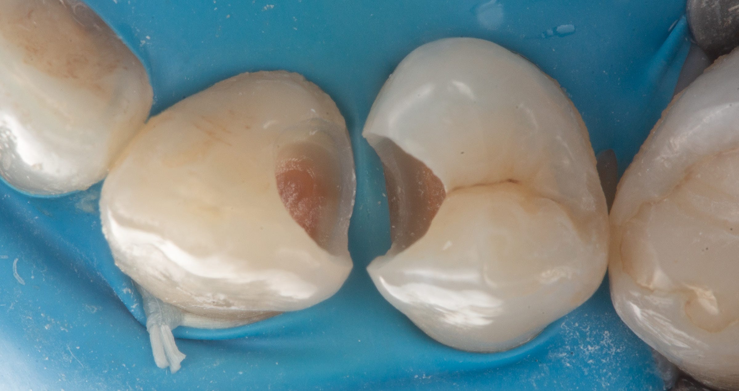
A possible clinical protocol is illustrated in Figures 2 to 5: After caries excavation and tooth preparation, sectional matrices, wedges and rings were placed to allow for simultaneous treatment of the mesial and occlusal cavities. Following etching and application of the universal adhesive CLEARFIL™ Universal Bond Quick (Kuraray Noritake Dental Inc.), the cavities were restored with CLEARFIL MAJESTY™ ES Flow Super Low in the shade A1 and CLEARFIL MAJESTY™ ES-2 Universal in the shade U. The distal cavity of the first molar was filled in the last step of the free-hand modeling procedure. In order to restore the occlusal surfaces in their original vertical dimension, every second tooth was isolated with rubber dam and the exposed molar etched (total-etch technique with K-ETCHANT Syringe, Kuraray Noritake Dental Inc.). the alternating index was positioned with some pressure and the flowable composite (CLEARFIL MAJESTY™ ES Flow Super Low) injected. Once light curing was completed, it was possible to remove the index, chip off the excess and finish and polish the restoration before repeating the procedure for the adjacent molar.


Fig. 2. Restoration of two molars: Teeth preparation and caries excavation.


Fig. 3. Restoration of two molars: Filling of the proximal and occlusal cavities.


Fig. 4.  Restoration of two molars: Re-establishing the occlusion with the aid of the flowable injection technique.


Fig. 5. Alternating approach: Restoration of the second molar by injecting flowable composite.


DISCUSSION

The use of the flowable injection technique allows for rapid restoration of teeth and the establishment of precise occlusal contacts. This reduces the time spend on occlusal surface modelling and minimizes the risk for prolonged treatment due to a repeated need for occlusal adjustments. In addition to saving time, it is possible with this technique to restore a greater number of teeth in a single appointment. The aesthetics of this type of restoration may be somewhat limited: A skilled practitioner is able to achieve better aesthetic results on the occlusal surface. However, with a detailed wax-up and high-quality model great outcomes can be obtained. The surface quality of printed models can be increased by adjusting the printing parameters including the layer height (Fig. 6). The use of a hydraulic pressure curing unit for silicone index production further increases the quality of the occlusal surface.


When planned and implemented correctly, the established occlusal surface and contacts reflect the natural anatomy without the need for adjustments (Fig. 7). Especially when restoring an entire quadrant, it is possible to increase the efficiency by opting for the flowable injection technique. Doing so reduces the number of appointments and the chair time decisively (Fig. 8).


STAMP TECHNIQUE: CONSIDERATIONS

If the occlusal surface of the tooth is intact, a wax-up may not be necessary. In this case, the better strategy is to duplicate what is still available before initiating treatment. A flowable composite or liquid rubber dam can be used for this purpose. It is important to coat the tooth surface with glycerin gel before applying the material. This will facilitate separation of the stamp from the tooth. It is always advisable to create a stamp that covers not only the details that need to be recorded and duplicated, but is extended over the cusps. This offers better stability in the restoration phase.


CLINICAL PROTOCOL

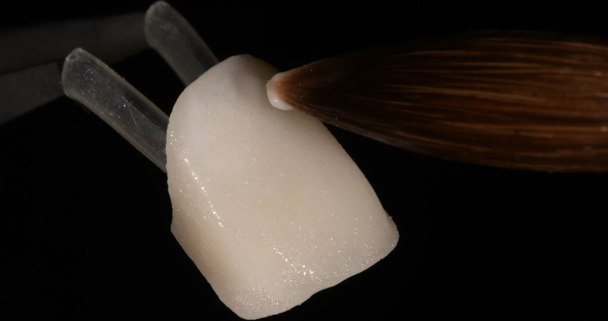
Figures 9 to 11 illustrate a possible clinical procedure. In this case, a molar with an occlusal carious lesion needed to be restored. The tooth surface was cleaned and a thin layer of glycerin gel applied, followed by a thick layer of liquid rubber dam, which covered the entire occlusal surface. Then, a micro applicator was immersed into the material and the stamp cured. After preparation, etching and application of the bonding system, the cavity was restored with flowable composite (CLEARFIL MAJESTY™ ES Flow Super Low in the shade A2). When the cavity is larger and depending on personal preferences, a paste-type composite (CLEARFIL MAJESTY™ ES-2 Universal) may also be used. Prior to light curing of the composite, the occlusal surface was covered with PTFE tape and the stamp pressed onto it. After firm pressing, the tape and excess material were removed and the restoration polymerized. This restoration faithfully reproduces the occlusal surface and did not require any occlusal adjustments.


Fig. 6. Stamp production with liquid rubber dam.


Fig. 7. The stamp.


Fig. 8. Restoration procedure: From preparation to bonding.


Fig. 9. Restoration procedure: Filling with flowable composite.


Fig. 10. Restoration procedure: Duplication the original occlusal surface with the stamp.


Fig. 11. Tooth before and after treatment using the stamp technique.


CONCLUSION

Techniques that add simplicity and efficiency to clinical procedures are always welcome in the busy practice environment. Depending on the information available at the start of treatment and the number of teeth to be restored, the flowable injection or the stamp technique may be an ideal choice. They are easily implemented and speed up the clinical procedure, but most importantly support predictable outcomes. This saves time in the finishing phase and minimized the risk of repeated adjustments, hence protecting everyone involved from additional appointments and frustration. Especially for practitioners with limited routine in free-hand modelling and for those with maximum patient comfort in mind, both techniques are worth being integrated in their clinical procedures.


Dentist:

MICHAL JACZEWSKI


Michał Jaczewski graduated from Wroclaw Medical University in 2006 and today runs his private practice in the city of Legnica, Poland. He specializes in minimally invasive dentistry and digital dentistry and is the founder of the Biofunctional School of Occlusion. Here he lectures and runs workshops with focus on full comprehensive patient treatments.


Zobu cirkonijs Iemesli, kādēļ zobārstiem vajadzētu iesaistīties protezēšanas materiālu izvēlē

Augstas kvalitātes protezēšanas nozīme

Augstas kvalitātes ārstēšana, iespējams, ir vissvarīgākais faktors ceļā uz pacienta apmierinātību. Katrā vizītē pacients vēlas justies kvalificēta speciālista aprūpēts. Savukārt krēslā pavadītais laiks un vizīšu skaits ir jāsamazina līdz nepieciešamajam minimumam. Tas nozīmē, ka protezēšanas kontekstā restaurācijai nekavējoties ir perfekti jāpieguļ un laika gaitā tai jābūt stabilai, lai izvairītos no pārtaisīšanas un papildus apmeklējumiem.


Bet kā iespējams katru reizi nodrošināt perfekti pieguļošas, kvalitatīvas restaurācijas? Starp potenciālajiem netiešo restaurāciju kvalitātes problēmu avotiem var minēt bieži pieļautās kļūdas zobārstniecības kabinetā vai laboratorijā, komunikācijas problēmas un bieži vien zemas kvalitātes zobu cirkonija oksīda izmantošanu.


Cirkonija restaurācijas – mūsdienīgs un estētisks zobārstniecības risinājums

Vairāk nekā pirms 20 gadiem cirkonija oksīds ienāca zobārstniecības tirgū kā kroņu un tiltu ražošanā izmantotā metāla aizstājējs. Abi materiāli – gan cirkonija oksīds, gan metāls – parasti tika apvienoti ar porcelāna slāni, veidojot porcelāna, metālā izkausēta porcelāna vai cirkonijā izkausēta porcelāna restaurācijas. Turpmākajos gados vairāki vadošie zobārstniecības cirkonija oksīda ražotāji (piemēram, Kuraray Noritake Dental Inc.) koncentrējās uz materiāla uzlabojumiem. Šie uzlabojumi pakāpeniski pārveidoja sākotnējo balti necaurspīdīgo karkasa materiālu keramikas materiālā ar zobiem līdzīgām optiskajām un izcilām mehāniskajām īpašībām. Daudzi zobārstniecības speciālisti visā pasaulē uzskata jaunākos cirkonija oksīda variantus, kas pieejami ar dažādiem caurspīdīguma un stiprības līmeņiem, par labāko iespējamo ārstēšanas iespēju dažādiem pacientiem un indikācijām. Viens no iemesliem ir tas, ka tiem ir nepieciešams tikai neliels porcelāna slānis, vai vispār nav vajadzīgs. Cits iemesls ir tas, ka ar mazu minimālo sieniņu biezumu tie ļauj veikt konservatīvu zobu sagatavošanu, vienlaikus nodrošinot labvēlīgu ilgtermiņa kalpošanu – ar noteikumu, ka tiek izmantots augstas kvalitātes materiāls.


Zobu cirkonija kvalitātes atšķirības

Cirkonija produktu kvalitāte var atšķirties atkarībā no dažādiem faktoriem, piemēram, izejvielu tīrības (ne tikai cirkonija, bet arī alumīnija oksīda un itrija, kā arī krāsvielu piedevu utt.), precīza ķīmiskā sastāva, daļiņu izmēra un sadalījuma. Katrs solis sagatavju ražošanas procesā – no pulvera sagatavošanas līdz sagatavju presēšanai un sinterizācijai sastiprināšanai – ietekmē gala kvalitāti, t.i., arī cirkonija mehāniskās un optiskās īpašības.


Bieži sastopamās problēmas, ko izraisa zemas kvalitātes cirkonija oksīds

Ikreiz, kad kaut kas nav kārtībā ar restaurācijas optiskajām īpašībām – ar tās caurspīdīgumu, kopējo krāsu vai pāreju no viena slāņa uz nākamo sagatavēs ar daudzslāņu krāsu struktūru – problēma kļūs acīmredzama pēc sinterizācijas laboratorijā. Rezultātā var būt nepieciešamība pārtaisīt, kā arī defekts var tikt atklāts laikošanas laikā, kas, visticamāk, negatīvi ietekmēs pacienta apmierinātību. Tas pats attiecas uz gadījumiem, kad, piemēram, materiāla struktūras neviendabīgums izraisa neprecīzu piegulēšanu. Vēl sliktāk ir zemākas pakāpes bioloģiskā saderība, virsmas kvalitāte, malu stabilitāte, lieces stiprība vai izturība pret lūzumiem. Šīs problēmas var identificēt tikai ar testēšanas aprīkojumu, kas ir ļoti dārgs un parasti nav pieejams zobārstniecības laboratorijās. Tas nozīmē, ka šāda veida nepilnības parasti paliek neatklātas līdz rodas reāla klīniska problēma, piemēram, smaganu recesija, palielināta aplikuma uzkrāšanās, lielāks nodilums vai agrīns defekts, kas var izraisīt sāpes un diskomfortu.


Overview of potential problems and clinical consequences for patients

LIespējamas ar nestandarta cirkoniju saistītas
problēmas

Lespējamās klīniskās sekas pacientiem

Ierobežota bioloģiskā saderība

Smaganu recesija / iekaisums

Neviendabīgums materiāla struktū

Neprecīza restaurācijas piegulēšana

virsmas plaisas

estētiskas problēmas (caurspīdīgums, krāsa) > atkārtota izveide

Zemāka virsmas kvalitāte: poraina virsma

Paaugstināta aplikuma uzkrāšanās > periodonta problēmas, kariess

Zemāka virsmas kvalitāte: raupjāka virsmas tekstūra

Grūtāk izlīdzināt un nopulēt > augsts antagonistu nodilums

Slikta malu stabilitāte

Malu plaisas un lūzumi > agrīna labošana vai nomaiņa

Zema lieces stiprība

Samazināta ilgmūžība > agrīna nomaiņa

Ierobežota izturība pret lūzumiem

Lūzumi / ierobežota ilgmūžība > agrīna nomaiņa


Zobu cirkonija sertifikācija un standartizācija

Speciālisti ir izstrādājuši ISO standartu (ISO 6872:2015), kurā aprakstīti in vitro testi, kas jāveic katram Eiropā vai ASV izmantotā zobārstniecības cirkonija ražotājam, lai saņemtu FDA apstiprinājumu un CE marķējumu. Aprakstītie testi tiek izmantoti, lai izmērītu lieces stiprību un izturību pret lūzumiem, kas, iespējams, ir divas vissvarīgākās īpašības, kas nosaka no materiāla izgatavoto restaurāciju ilglaicīgu darbību. Katram Eiropā vai Amerikas Savienotajās Valstīs izmantotajam materiālam ir jānokārto šie testi.


Kā izvairīties no zemas kvalitātes zobu cirkonija restaurāciju ievietošanas pacienta mutē

Ikvienam, kas izmanto šo sertificēto zobu cirkoniju, jābūt pārliecinātam un jāspēj samazināt ar materiāliem saistītos riskus. Tomēr zobu cirkonija pieaugošā popularitāte ir piesaistījusi to uzņēmumu uzmanību, kuri cenšas iegūt savu kumosa daļu, neveicot nepieciešamos pasākumus, lai nodrošinātu augstu produkta kvalitāti un nokārtotu sertifikāciju. Nesertificētiem produktiem, kuriem nav CE marķējuma, ir viena kopīga iezīme: tie noteikti rada draudus Jūsu uzņēmumam un pacientiem.

Ņemot vērā augstāk aprakstīto kā ir iespējams nodrošināt cirkonija produktu kvalitāti zobārstniecībā? Labā ziņa ir tāda, ka ir pieejami daži vienkārši noteikumi. Ievērojot tos, jūs varat izvairīties no viltotu vai zemas kvalitātes zobu cirkonija restaurāciju ievietošanas pacienta mutē.


Izvairieties no viltotu vai zemas kvalittes zobu cirkonija restaurciju ievietoanas pacienta mutē.


Trīs zelta likumi, lai nodrošinātu saviem pacientiem augstas kvalitātes cirkonija restaurācijas:

  • Pasūtiet tikai tādas restaurācijas, kas ražotas vietējā tirgū vai reģionā ar tādiem pašiem standartiem kā jums: piemēram, Ķīnas zobārstniecības laboratorijās ražotām restaurācijām ir jāatbilst zemākiem standartiem (tātad tām nav CE marķējuma), un tās var neatbilst jūsu cerībām.
  • Konsultējieties ar savu (vietējo) laboratoriju par viņu cirkonija oksīda ražotāju, pārliecinieties, vai viņi iegādājas cirkoniju no vadošajiem ražotājiem (piem., Kuraray Noritake Dental Inc.), izmantojot pilnvarotus izplatītājus vai pārdevējus, kurus viņi pazīst.
  • Izvairieties no piedāvājumiem, kas ir pārāk labi, lai būtu patiesi: zemas cenas var būt vilinošas, taču ārstēšanas galīgās izmaksas var būt pat augstākas nekā parasti, ja rodas komplikācijas.


Ilgtermiņa ietekme pacientiem, izmantojot sertificētas cirkonija restaurācijas

Pārliecība, ka jūsu zobārstniecības kabinetā izmantotais cirkonijs atbilst augstākajiem iespējamajiem kvalitātes standartiem, ir svarīgs ieguldījums pacientu ilgtermiņa apmierinātībā. Pat ja augstas kvalitātes cirkonija restaurācijas sākotnējās izmaksas ir nedaudz augstākas nekā zemākas kvalitātes darba izmaksas, kopējais ieguldījums var būt mazāks, ja restaurācija kalpo ilgāk un tiek novērsta pārtaisīšana. Jūsu apmierinātie pacienti, visticamāk, būs vairāk iesaistīti un ievēros mutes higiēnu, kā arī būs lojāli, tādējādi pozitīvi ietekmējot jūsu reputāciju un pacientu bāzi.


Izpētiet cirkonija iespējas un izvēlieties produktus no sertificētiem ražotājiem

Ja vēlaties spert soli tālāk, varat pat salīdzināt vairāku ražotāju sertificētos cirkonija variantus un atklāt atšķirības. Piemēram, Kuraray Noritake Dental Inc. ir viens no nedaudzajiem zobu cirkonija mražotājiem, kas veic visu ražošanas procesu, tostarp izejvielu ražošanu uz vietas. Tādā veidā uzņēmums spēj kontrolēt katru procedūras soli un nodrošināt izcilu produkta kvalitāti – neatkarīgi no tā, kurš materiāla variants tiek izvēlēts. Ar pieejamo sortimentu, kas sastāv no KATANA™ Zirconia UTML (īpaši caurspīdīgs daudzslāņu), KATANA™ Zirconia STML (izcili caurspīdīgs daudzslāņu) un ļoti caurspīdīga daudzslāņu HTML PLUS, kā arī YML (ar papildu stiprības un caurspīdīguma gradāciju), ir iespējams aptvert praktiski visas indikācijas.


Universālais sveķu cements: Vai kādreiz esat domājis par trešo uzklāšanas veidu?

PROF. LORENCO BREŠI (LORENZO BRESCHI) RAKSTS


Mazāk pudeļu, vairāk izvēles – tas, iespējams, ir īsākais veids, kā aprakstīt universālo sveķu cementu kategoriju. Šie divkāršās cietēšanas sveķu cementi, kas ir pašlīmējoši, nodrošina vienkomponenta darbplūsmu, daudzās klīniskās situācijās neizmantojot atsevišķus zobu vai restaurācijas praimerus. Šādā veidā iegūtā saķeres stiprība parasti ir pietiekami augsta, lai nodrošinātu stabilu saķeri starp zobu un restaurāciju plašā indikāciju diapazonā. Tomēr tā ir nedaudz zemāka nekā tā, kas tiek sasniegta ar parastajām sveķu cementa sistēmām, kas sastāv no vairākiem komponentiem (parasti zobu saite sveķu cements un restaurācijas saite).

Papildus pašlīmējošajam uzklāšanas veidam universālos sveķu cementus var kombinēt ar papildu sistēmas sastāvdaļām, lai attiecīgi palielinātu saķeres stiprību ar zoba struktūru vai restaurācijas materiālu. Tas paver jaunas iespējas produkta lietošanā: atkarībā no nepieciešamās vai vēlamās saķeres veiktspējas universālo sveķu cementu var uzklāt atsevišķi vai kombinācijā ar zobu saiti restaurācijas saiti vai abām sastāvdaļām. Turklāt hibrīda koncepcijas kļūst iespējamas, kā paskaidrots šajā rakstā, kurā galvenā uzmanība pievērsta PANAVIA™ SA Cement Universal (Kuraray Noritake Dental Inc.) kā piemēram.


Pašcementējošā fiksācija: daudzām indikācijām

PANAVIA™ SA Cement Universal ir divējādi cietējošs universāls sveķu cements, kas ir paredzēts plašam pielietojumu klāstam, ja to izmanto pašcementējošā veidā. Saķere, kas izveidota ar restaurācijas substrātiem (tostarp silikāta keramiku), ir spēcīga, neizmantojot atsevišķu saiti vai silānu1-4. Tas ir saistīts ar diviem dažādiem lipīgajiem monomēriem, ko satur preparāts – oriģinālais MDP monomērs un LCSi monomērs (garas oglekļa ķēdes silāna savienošanas līdzeklis, kas atbild par spēcīgu ķīmisko saiti ar silikāta keramiku). Tādējādi ir iespējams izmantot sveķu cementu bez papildu komponentiem, kas tiek uzklāti uz restaurācijas sāniem – pat gadījumos, kad trūkst saķeres un līdz ar to ir augstas prasības attiecībā uz saķeres stiprību.

Spēcīga saķere ar emalju un dentīnu tiek iegūta arī pašlīmējošā veidā. Tomēr dažās situācijās var būt lietderīgi vēl vairāk palielināt saķeres stiprību ar zoba struktūru, izmantojot zobu saiti.


Adhezīva līmēšana: sarežģītām situācijām

PANAVIA™ SA Cement Universal ieteicamā zobu saite ir CLEARFIL™ Universal Bond Quick (Kuraray Noritake Dental Inc.). Tā lietošana ir ieteicama ikreiz, kad lietotājs uzskata, ka ārstēšanai noderētu īpaši spēcīga un izturīga ķīmiskā saite, t.i., īpaši sarežģītās situācijās ar nepietiekamu mehānisko noturību. Šī pasākuma efektivitāte ir apstiprināta Japānā veiktā in vitro pētījumā, kurā 24 stundu mikro stiepes saites stiprība pret dentīnu tika ievērojami palielināta, uzklājot universālo cementu5. Izmantojot atsevišķu cementu pieaug pilnīgi sausa darba lauka nozīme. Iemesls ir tāds, ka sveķu cementa mitruma tolerance parasti ir augstāka nekā cementiem. Līdz ar to koferdama uzlikšana ir ļoti ieteicama.


Selektīva adhezīva cementēšana: īsiem noslīpētiem zobiem un subgingivālajām malām

Situācijās, kad ir grūti pareizi izolēt darba lauku ar koferdamu ir pieejama trešā uzklāšanas iespēja, ko piedāvā Itālijas pētnieku grupa: selektīva adhezīva fi ksācija. Šajā gadījumā CLEARFIL™ Universal Bond Quick tiek uzklāts tikai uz tām sagatavotā zoba daļām, kas nodrošina pareizu mitruma kontroli, vienlaikus paļaujoties uz PANAVIA™ SA Cement Universal pašcementējošo funkcionalitāti vietās, kur ir grūti iegūt vēlamo sauso darba lauku. Situācijas, kas ir paredzētas šai tehnikai, ir slīpēti zobi ar subgingivālu sagatavošanu un īpaši īsi slīpēti zobi (kas kavē koferdama novietošanu).


Selektīvās adhezīva fiksācijas tehnikas efektivitāte ir pārbaudīta in vitro pētījumā, kurā tika salīdzinātas trīs līmēšanas stratēģijas – pašcementējošā fiksācija pilnībā adhezīva fiksācija un selektīva adhezīva fiksācija – ar bīdes saišu stiprības pārbaudes palīdzību6. Pārbaužu rezultāti liecina, ka lietotāji var uzlabot PANAVIA™ SA Cement Universal saķeres stiprību ar dentīnu un emalju, uzklājot cementu tikai uz zoba virsmas daļas. Cementēšanas sistēmai, kas sastāv no PANAVIA™ SA Cement Universal un CLEARFIL™ Universal Bond Quick, pilnībā adhezīva un selektīvā adhezīva pieeja radīja līdzīgus rezultātus.


Situācij ās, kad ir grūti pareizi izolēt darba lauku ar koferdamu ir pieejama trešā uzklāšanas metode ko piedāvā Itālij as pētnieku grupa: selektīva adhezīva fiksācija


Rekomendētās darbības selektīvajai adhezīva līmēšanai

1. attēls. Zoba sagatavošana


2. attēls. Selektiva emaljas kodinašana ar fosforskabes kodinataju


3. attēls. Universala adheziva uzklašana un žavešana ar gaisu


4. attēls. Krona novietošana pec sveku cementa uzklašanas kroni


5. attēls. Cietinašana


6. attēls. Lieka daudzuma noemšana un galeja cietinašana
gaisma


7. attēls. Arstešanas rezultats pec viena gada


Selektīvās adhezīva cementēšanas priekšrocības

Papildus vēlamajam (ilgtermiņa) saķeres stiprības pieaugumam, kas tiek panākts, uzklājot atsevišķu cementu uz daļas vai visas sagatavotās zoba virsmas, tehnika piedāvā papildu priekšrocības. Salīdzinot ar daudzpakāpju cementēšanas sistēmām, protokols ir vienkāršots, jo nav nepieciešams atsevišķa restaurācijas saite. Cementa cietināšana gaismā nav nepieciešama, kamēr lietotājs paliek ieteiktajā sistēmā. Un atšķirībā no pilnībā adhezīvas pieejas, kurā ir nepieciešams uzstādīt koferdamu šī soļa nepieciešamība ir izslēgta, izmantojot selektīvo adhezīva pieeju. Tādā veidā tiek samazināts krēslā pavadītais laiks un palielināts pacienta komforts.


Secinājumi

Atkarībā no indikācijas, klīniskajiem mainīgajiem lielumiem un individuālajām vēlmēm, universālo sveķu cementa, piemēram, PANAVIA™ SA Cement Universal, lietotāji var izvēlēties tehniku, kas, visticamāk, nodrošinās vislabākos klīniskos rezultātus. Tieši šī elastība un kopumā plašais pielietojumu klāsts padara inovatīvo produktu kategoriju patiesi universālu. Tā kā izmantojamo komponentu skaits ir mazāks, universālie materiāli atvieglo klīnisko procedūru racionalizāciju un standartizāciju, savukārt ar mazāku uzglabājamo pudeļu skaitu tas palīdz darbiniekiem iegūt kontroli pār pasūtījumu un uzglabāšanas pārvaldību.


Dentist:

LORENZO BRESCHI


Prof. Lorenzo Breschi ir restauraciju un zobarstniecibas materialu profesors Bolonas Universitate. Vinš aktivi iesaistas emaljas un dentina ultrastrukturalo
aspektu petijumos. Vinš ir bijušais Zobarstniecibas materialu akademijas (ADM) prezidents, Eiropas Konservativas zobarstniecibas federacijas (EFCD) ieveletais prezidents, Zobarstniecibas materialu grupas IADR ieveletais prezidents, Italijas Konservativas zobarstniecibas akademijas (AIC) ieveletais prezidents, Starptautiskas adhezivas zobarstniecibas akademijas (IAAD) ieveletais prezidents.


Atsauces

1. Cowen M, Cunha S, Powers JM. Novel Cement Bond Strength to Multiple Substrates.
DENTAL ADVISOR Biomaterials Research Center, Biomaterials Research Report, Number
132 - June 16, 2020. 2. Patel N, Anadioti E, Conejo J, Ozer F, Mante F, Blatz M. Bond
Strength of Different Self-Adhesive Resin Cements to Zirconia” (2021). Dental Theses.
62. https://repository.upenn.edu/dental_theses/62 3. Yoshihara K, Nagaoka N, Maruo Y,
Nishigawa G, Yoshida Y, Van Meerbeek B. Silane-coupling effect of a silane-containing
self-adhesive composite cement. Dent Mater. 2020 Jul;36(7):914-926. 4. Irie M, Tokunaga
E, Maruo Y, Nishigawa G, Yoshihara K, Nagaoka N, Minagi S, Matsumoto T. Shear bond
strength of a resin cement to CAD/CAM Blocks for molars. P-2, 37th Annual Meeting of the
Japanese Society of Adhesive Dentistry 2018. 5. Ohara N. Bonding strength of resin cement
containing silane coupling agent to dentin or core resin. Results presented at the 150th
meeting of the Japanese Society of Conservative Dentistry. 6. Breschi L, Josic U, Maravic
T, et al. Selective adhesive luting: A novel technique for improving adhesion achieved by
universal resin cements. J Esthet Restor Dent. 2023;1-9. doi:10.1111/jerd.13037


A GUIDE TO SUCCESSFUL ZIRCONIA BONDING


Unlock the power of zirconia: perfect for adhesive cementation, the ideal material for a wide range of indications, and essential in minimal invasive dentistry. Time to trust zirconia bonding!

This article demystifies zirconia bonding, providing clear, practical steps to ensure long-term functionality and patient satisfaction, all based on scientific research. Master the three adhesion pillars: mechanical retention, chemical activation, and wetting capacity. Discover how to successfully prepare zirconia surfaces, avoid pitfalls like misapplying silica coating and silane, and choose proven bonding systems for optimal results. Optimise retention even with minimal tooth preparation and achieve reliable zirconia restorations. Say goodbye to doubts and hello to successful zirconia bonding!


Factors influencing retention

Loss of retention due to de-cementation or debonding is a common cause of dental prostheses' failure.  First, let’s have a look at how to cope with the three main factors significantly influencing retention: tooth preparation, restoration pre-treatment, and cement type/bonding.

Tooth preparation

The abutment tooth's height, angle, and surface texture must be considered to achieve sufficient retention and resistance from the preparation. The retention form counteracts tensile stresses, whereas the resistance counteracts shear stresses 4. With the proper preparation, a restoration resists dislodgement and subsequent loss.

Full coverage restorations

To achieve sufficient retention and resistance for full-coverage crowns, the tooth abutment should be at least 4 mm high, and the convergence angle should range from 6 to 12 degrees with a maximum of 15 degrees 1, 5-8.

Source; Conventional cementation or adhesive luting - A guideline, Dr. A. Elsayed, Prof. Dr Florian Beuer 


Adhering to the tooth preparation guidelines is crucial for full-coverage restorations (e.g., crowns, and FDPs). These practical guidelines are designed to achieve the required retention and resistance to make conventional luting possible. However, optimal retention and resistance are, in reality, hard to achieve. An unwanted amount of sound tooth substance often should be removed to achieve a highly retentive preparation. Moreover, several studies2,3 show that, in daily practice, the preparation angle often exceeds 15 degrees.

Minimal-invasive restorations

Minimal-invasive restorations, such as single retainer FDPs, veneers, table-tops and inlay-retained FDPs, are based on a non- or low-retentive preparation form. In this case, retention shifts from (macro-)mechanical to micro-mechanical and chemical, necessitating the use of adhesive techniques 9-11. Even though the preparations for minimal-invasive restorations largely lack mechanical retention, the long-term success of these types of restorations is well-documented when using a suitable resin cement (e.g. PANAVIA™, Kuraray Noritake Dental, Japan), including a proper pre-treatment and bonding procedure 10, 11.

In high-retentive situations, conventional luting is acceptable for full-coverage restorations*. In all other cases, choosing a resin cement is a better solution. With proper tooth preparation (e.g., shaping, (self-)etching, abrasion) and the right adhesive resin cement system, a non-retentive preparation form provides a reliable basis using mainly chemical retention and micro-mechanical retention instead of macro-mechanical retention.

*Please review the articles available regarding the debate over whether to use a conventional cementation procedure, adhesive cementing, or selective adhesive luting


Restoration pre-treatment

Zirconia is densely sintered and does not contain a glass phase. Therefore, it cannot be etched with hydrofluoric acid to create a micro-retentive etching pattern. In addition, silanes cannot effectively promote zirconia bonding. Several studies have shown that air abrasion with 50-µm alumina at a reduced pressure of 0.5 bar (0.05 MPa; 7 psi) will create a sufficient micro-retentive pattern12 and greatly enhances the wetting capacity.

In addition to air abrasion, chemical coupling agents such as bifunctional phosphate resin monomers are used on air-abraded zirconia. Bonding with phosphate monomer-containing adhesive resin systems gives very reliable results27,28. The use of phosphate monomer-based resin cement systems (e.g., Panavia [Kuraray Noritake Dental, Tokyo, Japan]) and/or phosphate monomer primers, such as CLEARFIL CERAMIC Primer Plus (Kuraray Noritake Dental, Tokyo, Japan)  on freshly air-abraded zirconia, offer the most reliable bonding methods today 13,27,28. We therefor consider MDP-based composite resin cements the material choice for our bonding procedure. However, it must be stressed that contamination of the air-abraded zirconia with saliva, phosphoric acid or other contaminants will limit the formation of chemical bonds and, therefore, must be avoided.

Avoiding contamination

For optimal moisture control, absolute isolation of the working field is crucial. Minimising  the risk of contamination, avoiding exposure to oral fluids. Before restoration placement, a thorough cleaning of the abutment tooth is essential. Following trial placement, a meticulous recleaning step is recommended to remove any potential introduced contamination. KATANA Cleaner (Kuraray Noritake Dental, Tokyo, Japan) is an ideal choice due to its unique properties. Its slightly acidic pH of 4.5 allows for effective cleaning intraoral and extraoral adhesion surfaces. Additionally, the incorporation of MDP monomer technology makes it highly efficient. The MDP salt in this product effectively bonds with contaminants, breaks them down and results in easy removal by water rinsing.


Cement type/bonding

After pre-treatment of surfaces to optimise the , it is important to understand that the properties of highly translucent zirconia differ highly from those of earlier generation zirconia. Early-generation zirconium oxides, including 3 mol% yttrium oxide (3Y-TZP), are high in strength and low in translucency. With the increase in yttria, creating 4-5 mol% yttria, or higher, zirconium oxides, the number of cubic crystals increases, resulting in higher translucency but leading to a reduction in strength. Therefore, attention must be paid to zirconia type, material thickness, restoration type, and application area. These factors may influence the choice of cement based on the adhesive properties demanded for lasting restorations and high aesthetic outcomes.


PANAVIA V5

For a resin cement system to deliver a strong bond, it is not always enough to have it contain an appropriate adhesive monomer. It is necessary for that adhesive monomer to be polymerised effectively under different circumstances. The PANAVIA™ V5 system contains an innovative “ternary catalytic system” consisting of a highly stable peroxide, a non-amine reducing agent* and a highly active polymerisation accelerator. Since this catalytic system is amine-free, the hardened cement has unsurpassed colour stability. In addition, the highly active polymerisation accelerator, one of the components in PANAVIA™ V5 Tooth Primer, is not only an excellent reducer that promotes polymerisation effectively, but it is also capable of coexisting with the (in this product) acidic MDP. This makes it possible to create a single-bottle self-etching primer. This accelerator is also responsible for the so-called touch-cure reaction when it comes into contact with the paste. Resulting in the sealing of the dentin interface and, at the same time, allowing the paste to set even in situations where light curing is limited.

*PANAVIA™ V5 Tooth Primer applied and left for 20 seconds, followed by air drying.

The second primer in the PANAVIA V5 system is CLEARFIL™ CERAMIC PRIMER PLUS, which incorporates Kuraray Noritake Dental’s original MDP and a silane. This product is used to prime zirconia but is also an excellent choice for priming silica-based ceramics, composites, and metals.


CLEARFIL™ CERAMIC PRIMER PLUS, which contains the original MDP, applied and dried.


The PANAVIA™ V5 full adhesive resin cement system consists of all three above-mentioned components, always used in the same way, independent of the material, for a straightforward procedure to ensure reliable bonding. The PANAVIATM V5 systems offer try-in pastes to visualise the final results before final cementing and confirm the appropriate shade of the resin cement to be used.


PANAVIA VENEER LC


Offering a flexible workflow and high bondability of thin, translucent restorations like veneers but also inlays and onlays, PANAVIA™ Veneer LC was designed. It is a light-curing resin cement system allowing a long working time of 200 seconds under ambient light*. This allows multiple veneers to be placed simultaneously without racing against the setting. The final light-curing can be started anytime after positioning the provisions. The PANAVIA™ Veneer LC cementing system includes PANAVIA™ Tooth Primer and CLEARFIL CERAMIC PRIMER Plus as primers to chemically interact with the adhesive surfaces.


PANAVIA™ Veneer LC Paste applied and the laminate veneer seated. In this case six veneers were simultaneously placed during one session.

Unpolymerized excess paste removed with a brush. PANAVIA™ Veneer LC Paste is a light-cured type rein cement, designed to provide sufficient working time.

This photo shows the results after the final light curing. Since the excess cement was easily removed, there were almost no cement residues.


PANAVIA SA CEMENT Universal

Still, clinicians seek efficiency and effectiveness in everyday practice by using a straightforward but durable resin cement solution. PANAVIA™ SA Cement Universal is developed to offer this ease-of-use property without losing focus on bonding properties.  PANAVIA™ SA Cement Universal is developed with the original MDP monomer in the hydrophilic paste compartment, allowing for chemical reactiveness with zirconia and tooth structure. The other compartment contains the hydrophobic paste, to which a unique silane coupling agent, LCSi monomer, is added, which allows the cement to deliver a strong and durable chemical bond to silica-based materials like porcelain, lithium disilicate and composite resin*. Furthermore, PANAVIA™ SA Cement Universal is less moisture sensitive than full adhesive resin cement systems. This also makes it the ideal cement in situations where rubberdam isolation is difficult.

*The product is available in both auto mix and hand mix options.

*Old PFM bridge (shown here) removed, and existing preparations modified to accommodate a 3-unit KATANA™ Zirconia bridge. The upper right canine was prepared to receive a single-unit KATANA™ crown.

Before

After. Seating & Final Smile. PANAVIA™ SA Cement Universal and CLEARFIL™ Universal Bond Quick were used for cementation and bonding. “I love the ease of use and clean-up with PANAVIA™ SA Cement Universal, and its MDP monomer creates a strong chemical bond to the tooth structure and zirconia. CLEARFIL™ Universal Bond Quick has a quick technique without reducing bond strengths, releases fluoride and has a low film thickness. I simply rub CLEARFIL™ Universal Bond Quick into the tooth for a few seconds and air dry. There is no need to light-cure, since it cures very well with PANAVIA™ SA Cement Universal. The patient was very happy with the results. She loved that she no longer saw metal margins, and her smile was much more uniform and lifelike.” Dr. Kristine Aadland


*Images are a part of a case by Dr. Kristine Aadland; 3-Unit anterior maxillary


Bonding to zirconia in three steps

Over the last century, the popularity of highly translucent zirconia has skyrocketed due to its excellent properties and wide range of anterior and posterior clinical applications. Because zirconium oxide prostheses are, if processed correctly, antagonist-friendly and easy (and relatively inexpensive) to fabricate, the material keeps gaining popularity in dentistry.

Several steps need to be taken into account for reliable and durable bonding. Years of research on achieving high and long-term bond strength to zirconia have concluded into three practical steps, summarised as the APC concept13 as a reliable procedure guideline.

APC-Step A

Zirconia should be air-particle abraded (APC-Step A) with alumina or silica-coated alumina particles; the sandblasting or micro-etching procedure. Air abrasion with a chairside micro-etcher using aluminium oxide particles (size: up to 50 μm) at a low pressure of 0,5 bar (0.05 – 0.25 MPa) is sufficient.14,18,25-27

APC-Step P

The subsequent step includes applying a special ceramic primer (APC-Step P), which typically contains specially designed adhesive phosphate monomers, onto the zirconia adhesive surfaces.29,30 The MDP monomer has been shown to be particularly effective at bonding to metal oxides like zirconium oxide.

APC-Step C

Dual- or self-cure resin cement systems should be used to reach an adequate C=C conversion rate underneath the zirconia restoration since the lack of translucency in zirconia reduces light transmission.13 However, in cases where high-translucent zirconia (HTZr02) is used, the zirconia transmits light so that the shade of composite or resin cement might influence the final appearance of such restorations. It is, thereforebased on the individual situation and shade of the abutment tooth.

The APC zirconia-bonding concept is not limited to intra-oral situations and can also be applied in the laboratory for implant reconstructions that include cemented zirconia components.

Conclusion

Rapid developments in high-quality translucent zirconia have made the utility and reliability of adhesive cementing systems even more crucial. This applies to fully opaque restorations but also minimally invasive and ultra-translucent restorations of low thickness. In all cases, the longevity of the bonding and, thus, the provision directly affects patient  satisfaction. By taking into account the three primary parameters we have discussed in this article and following the predictable APC protocol, you will successfully realise durable bonded zirconia restorations from now on.


References

  1. Ladha K, Verma M. Conventional and contemporary luting cements: an overview. J Indian Prosthodont Soc. 2010;10(2):79-88.

  2. Nam, Y., Eo, M.Y. & Kim, S.M. Development of a dental handpiece angle correction device. BioMed Eng OnLine17, 173 (2018). https://doi.org/10.1186/s12938-018-0606-1
  1. Florian BEUER, Daniel EDELHOFF, Wolfgang GERNET, Michael NAUMANN, Effect of preparation angles on the precision of zirconia crown copings fabricated by CAD/CAM system, Dental Materials Journal, 2008, Volume 27, Issue 6, Pages 814-820
  1. Muruppel AM, Thomas J, Saratchandran S, Nair D, Gladstone S, Rajeev MM. Assessment of Retention and Resistance Form of Tooth Preparations for All Ceramic Restorations using Digital Imaging Technique. J Contemp Dent Pract. 2018;19(2):143-9.

  2. Edelhoff D, Özcan M. To what extent does the longevity of fixed dental prostheses depend on the function of the cement? Working Group 4 materials: cementation. Clin Oral Implants Res. 2007;18 Suppl 3:193-204.

  3. Güth JF, Stawarczyk B, Edelhoff D, Liebermann A. Zirconia and its novel compositions: What do clinicians need to know? Quintessence Int. 2019;50(7):512-20.

  4. Smith CT, Gary JJ, Conkin JE, Franks HL. Effective taper criterion for the full veneer crown preparation in preclinical prosthodontics. J Prosthodont. 1999;8(3):196-200.

  5. Uy JN, Neo JC, Chan SH. The effect of tooth and foundation restoration heights on the load fatigue performance of cast crowns. J Prosthet Dent. 2010;104(5):318-24.

  6. Blatz MB, Vonderheide M, Conejo J. The Effect of Resin Bonding on Long-Term Success of High-Strength Ceramics. J Dent Res. 2018;97(2):132-9.

  7. Chaar MS, Kern M. Five-year clinical outcome of posterior zirconia ceramic inlay-retained FDPs with a modified design. J Dent. 2015;43(12):1411-5.

  8. Kern M, Passia N, Sasse M, Yazigi C. Ten-year outcome of zirconia ceramic cantilever resin-bonded fixed dental prostheses and the influence of the reasons for missing incisors. J Dent. 2017;65:51-5.
  1. Kern M, Dr Med Habil, M. BONDING TO ZIRCONIA. Jerd_40. 3DOI 10.1111/j.1708-8240.2011.00403.x VOLUME 2 3 , NUMBER 2 , 2011
  1. Blatz MB, Alvarez M, Sawyer K, Brindis M. How to Bond Zirconia: The APC Concept. Compend Contin Educ Dent. 2016 Oct;37(9):611-617; quiz 618. PMID: 27700128.
  1. Blatz M.B., Oppes S., Chiche G., et al. Influence of cementation technique on fracture strength and leakage of alumina all-ceramic crowns after cycling loading. Quintessence Int. 2008; 39(1): 23-32
  1. Burke F.J., Fleming G.J., Nathanson D., Marquis P.M. Are adhesive technologies needed to support ceramics? An assessment of the current evidence. J Adhes Dent. 2002;4(1)): 7-22
  1. Blatz M.B. Sadan A., Maltezos C., et al. In vitro durability of the resin bond to feldspathic ceramics. AM J Dent 2004;17 (3):169-172
  1. Blatz M.B., Bergler M. Clinical applications of a new self-adhesive resin cement for zirconium-oxide ceramic crowns. Compend Contin Educ Dent. 2012;33(10):776-781
  1. Maggio M., Bergler M., Kerrigan D., Blatz M.D. Treatment of maxillary lateral incisor agenesis with zirconia-based all-ceramic resin bonded fixed partial dentures: a case report. Amer J esthet Dent. 2012;2(4):226-237
  2. Ozer F., Blatz M.B., Self-etch and etch-and0rinse adhesive systems in clinical dentistry. Compend Contin Edus Dent. 2013;24 (1):12-20
  1. Kern M., Thomson V.P., Bonding to glass infiltrated alumina ceramic: adhesive methods and their durability. J Prosthet Dent. 1995;73 (3):240-249
  1. Kern M., Wegner S.M., Bonding to zirconia ceramics: adhesion methods and their durability. Dent Mater. 1998;14(1):64-71
  1. Wegner S.M., Kern M. Long-term resin bond strength to zirconia ceramic. J Adhes Dent. 2000;2 (2):139-147
  1. Blatz M.B., Sadan A., Martin J., Lang B. In vitro evaluation of shear bond strength of resin to densely-sintered high-purity zirconium-oxide ceramics after long-term sorage and thermos cycling. J Posthet Dent. 2004;9(4):356-362
  1. Blatz M.B., Chiche G., Holst S., Sadan A. Influence of surface treatment and simulated aging on bond strength of luting agents to zirconia. Quintessence Int. 2007;38 (9):745-753
  1. Quaas A.C., Yang B., Kern M., Panavia F 2.0 bonding to contaminated zirconia ceramic after different cleaning procedures. Dent Mater. 2007;23(4):506-512
  1. Song J.Y., Park S.w., Lee K., et al. Fracture strength and microstructire of Y-TZP zirconia after different surface treatments. J Prosthet Dent. 2013;110(4):274-280
  1. Koizumi H., Nakayama D., Komine F., et al. Bonding of resin-based luting cements to zirconia with and without the use of ceramic priming agent. J adhes Dent. 2012;14(4):385-392
  1. Nakayama D., Koizumi H., Komine F., et al. Adhesive bonding of zirconia with single -liquid acidic primers and a tri-n0butylborane initiated acrylic resin. J Adhes Dent. 2010;12(4):305-310
  1. Alnassar T., Ozer F., Chiche G., Blatz M.B. Effect of different ceramic primers on shear bond strength of resin-modified glass ionomer cement to zirconia. J Adhes Sci Technol. 2016;DOI:10.1080/01694243.1184404
  1. Blatz M.B. Long-term clinical success of all-ceramic posterior restorations. Quintessence Int. 2002;33(6):415-426
  1. Mante F.K., Ozer F., Walter R., et al. The current state of adhesive dentistry: a guide for clinical practice. Compend Contin Educ Dent. 2013;34:Spec 9:2-8
  1. Ozcan M., Bernasconi M. Adhesion to zirconia used for dental restorations: a systematic review and meta-analysis. J Adhes Dent. 2015;17(1):7-26
  1. Inokoshi M., De Munck J., Minakuchi S., Van Meerbeek B. Meta-analysis of bonding effectivenss to zirconia ceramics. J Dent Res. 2014;93(4):329-334


Tripartite talk

Presented by Kuraray Noritake Dental Inc.


Highly translucent multi-layered zirconia developed by a proprietary material and manufacturing method from Japan


CURRENT STATUS AND FUTURE PROSPECTS OF ZIRCONIA RESTORATIONS


In this issue, we asked Markus B. Blatz, Professor at the University of Pennsylvania, USA, Aki Yoshida (Gnathos Dental Studio) and Naoki Hayashi (Ultimate Styles Dental Laboratory), both dental technicians active in the USA and international instructors for Kuraray Noritake Dental Inc., to give their views on zirconia restorations and their outlook for the future.


WITH THE INTRODUCTION OF ZIRCONIA, THE MAINSTREAM OF PROSTHETIC TREATMENT HAS SHIFTED FROM METAL CERAMICS1 TO ZIRCONIA CERAMICS2. WHAT CHANGES HAVE OCCURRED WITH THE INTRODUCTION OF ZIRCONIA?


Blatz: My mentor for my first Ph.D. in dental materials was in the group that developed lithium disilicate and glass-infiltrated alumina. Therefore, I have seen the evolution of dental ceramic materials, including zirconia, which is the subject of this presentation, up close and personal.


Early zirconia was white, opaque, and not as esthetic as today. However, there is no doubt that zirconia ceramics were much more esthetic than metal ceramics. At the same time, however, we often heard the opinion that bilayer zirconia ceramic restorations were problematic, and this provoked much discussion. We conducted a large study in collaboration with a Boston laboratory to compare more than 1,000 posterior porcelain-fused-to-metal crowns and 1,100 posterior porcelain-fused-to-zirconia crowns and found no difference in chipping or fracture rates after about seven years. This proves that bilayer zirconia ceramics are safe when used with the proper veneering materials and the proper sintering and cooling protocols. The fact that zirconia became established as it is today is a major change for dentistry in general.

Yoshida: I also switched from metal ceramics to zirconia ceramics, and now I don't use metal anymore. It used to take a lot of time and effort to invest and cast metal, observe it with a microscope, and fit it. Considering the recent rise in metal prices, it has also become more cost-effective. In addition, I am allergic to metal and have a skin rash every time I have a prosthetic processed, so the shift to zirconia ceramics as the mainstream prosthetic is a welcome change. Of course, the use of zirconia has also improved esthetics. The translucency of zirconia is the greatest advantage that metal does not have.


Hayashi: Yes, that's right. The big advantage of zirconia is that if the abutment is not strongly discolored, it no longer needs to be treated with an opaquer. It was not easy to control the reflection of light from the operative tooth when fabricating metal ceramics. In addition to the esthetic advantage, the prosthetic space can be thinner than that of metal ceramics.


1. Metal ceramics: Prosthetic made of metal frame with porcelain.
2. Zirconia ceramics: Prosthetic made of zirconia frame with porcelain.


THE YEAR 2023 MARKED THE 10TH ANNIVERSARY OF THE FIRST MULTI-LAYERED ZIRCONIA – KATANA™ ZIRCONIA ML. SINCE THEN, HOW DO YOU THINK HIGHLY TRANSLUCENT MULTI-LAYERED ZIRCONIA HAS REVOLUTIONIZED PROSTHETIC DEVICE MANUFACTURING?


Yoshida: I feel the ability to extend the zirconia frame to the occlusal surface and the incisal edge is the greatest advantage of using highly translucent multilayered zirconia. This allows us to provide crowns of both esthetics and strength, even for patients with para function. I have also made a zirconia Maryland bridge using highly translucent multi-layered zirconia, and it is doing very well. There are some cases where it is not possible to use zirconia, but still, it is wonderful to have a wider range of options.


Blatz: Many people still have the impression that zirconia cannot be bonded to tooth structure, but resin cement can be used to bond zirconia to tooth structure after proper pretreatment. Clinical studies of resin-bonded zirconia bridges have shown very high success after 10 or 15 years. Currently, resin bonding is recommended for very thin, highly translucent zirconia, rather than cementation. However, it should be added that this requires the dentist and technician to understand the proper bonding technique for zirconia.


In addition, Kuraray Noritake Dental's multi-layered zirconia has revolutionized monolithic zirconia without the need for veneering porcelain. However, this has also resulted in the need for dental technicians to shift to a different approach: instead of building up the restoration as with veneering ceramics, esthetic features are created on the outer surface in each case.


Maxillary 6 anterior monolithic crowns (Markus B. Blatz)


Fig. 1a and b: Initial examination.


Fig. 1c: Simulation of final prosthetic restoration.


Fig. 1d: Completed prosthetic on model (monolithic crown using KATANA™ Zirconia STML).


Fig. 1e and f: Final restoration (Dr. Julian Conejo and Sean Han, CDT).


Two cases of Maryland bridge and laminate veneers and a mandibular canine single crown implant superstructure (Aki Yoshida)


Fig. 2a and b: Case 1: A case of a congenital defect of a lateral incisor was restored with a Maryland bridge. Since the proximal and distal width of the defect was greater than the central incisor, a non-prep veneer was fabricated on the central incisor to balance the proportions. KATANA™ Zirconia STML was used for the Maryland bridge. Note the harmony between the zirconia frame extended to the incisal edge and the transparency of the laminate veneers made of Super Porcelain EX-3™ on the central incisors. This case demonstrates the characteristics of zirconia, which combines strength and esthetics.


Fig. 3a to c: Case 2: A case of a screw-retained crown restoration of an implant placed in a mandibular canine tooth. Extension of the zirconia frame from the entire lingual side to the incisal margin prevents fracture of the porcelain by the screw access hole edges and canine guides. KATANA™ Zirconia STML provides natural transparency even when zirconia is exposed at the incisal edge.


Maxillary 4 Anterior teeth implant bridge (Naoki Hayashi)


Fig. 4a to f: Implant bridge of maxillary four anterior teeth using implants placed in the maxillary bilaterallateral incisors as abutments and maxillary bilateral central incisorsaspontics. The lingual side is fully backed with zirconia and the labial side is minimally layered with CERABIEN™ ZR.


Hayashi: Indeed, the highly translucent multilayered zirconia has expanded the possibilities of monolithic crowns. For patients with high occlusal forces, monolithic crowns are suitable in terms of strength, and with the use of highly translucent multilayered zirconia, it is possible to achieve a certain level of esthetics with monolithic crowns. In fact, some patients are happy with it. However, at least in the current situation, we believe that if patients and dentists want high-end esthetics, then porcelain buildup is necessary, and monolithic crowns are only an option.


Blatz: The variety of options available is the advantage of zirconia. The dentist and the technician can work together to provide the best possible outcome for the patient.


Yoshida: In terms of options, Kuraray Noritake Dental's zirconia can be sintered in a short time (approximately 90 minutes) in addition to the normal sintering time (7 hours) using a zirconia raw material and manufacturing method developed by Kuraray Noritake Dental, which is an advantage in that it can be used for immediate restorations, remanufacturing and other unexpected situations.


FINALLY, DO YOU HAVE A MESSAGE FOR THE NEW GENERATION OF DENTISTS AND DENTAL TECHNICIANS?


Blatz: I encourage my students and colleagues to always do their best. This leads to good results, makes you happy, and makes you feel satisfied with your life. Some people only try to get rich, but just accumulating wealth is never happiness. The second is to keep an open mind. Nowadays, we are inundated with information through social media.


Some of it is very stimulating and wonderful, but there is also a lot of it that is wrong. On the other hand, there are those who believe that everything one leader says must be done. I would like to tell them, "Make sure you get your information from reliable sources, and then choose reliable information for yourself. Dentistry is changing, so let's keep an open mind. The most important thing is that the patient is ultimately satisfied with the results.


Hayashi: I would like the future generation to learn more about tooth morphology, occlusion, and fit. Color is the essence of the quality of the final prosthetic device, but we need to learn tooth morphology, occlusion, and fit before we learn color. We are all about creating a prosthetic device that will function in the patient's mouth for the long term, and that is our goal. There will be new technologies and materials in the future, but their essence will never change. I hope that you will always remember what is important in your clinical practice. This is why basic knowledge of anatomy and function is necessary.


Yoshida: New technologies and materials will continue to emerge. But human teeth will not change. The most important thing is to provide the best possible care to the patient. I hope that you will accumulate such experiences, and that when you reach the end of your life, you will be able to say that you are glad you chose this profession.


Thank you very much for the meaningful discussion today.


Source: QDT Vol.49/2024 April
The magazine may not be printed from the web and may not be forwarded
No reproduction or reprinting allowed


Dentists:

Prof. Dr. Markus B. Blatz

University of Pennsylvania
School of Dental Medicine
240 S 40th St, Philadelphia,
PA 19104, USA

Aki Yoshida, RDT

Gnathos Dental Studio
56 Colpitts Rd, Weston,
MA 02493, USA

Naoki Hayashi, RDT

Ultimate Styles
Dental Laboratory
23 Mauchly Suite 111, Irvine,
CA 92618, USA


Empower your dental lab with KATANA Zirconia YML

KATANA™ Zirconia YML offers an unmatched blend of aesthetics and mechanical properties, but also provides for cost and time efficiencies.


Recognised for its strength and density at point of manufacture, the material delivers incredible hardness in its green state. This offers the fully validated opportunity to make adjustments in morphology directly after milling.


These qualities, along with its strength and translucency once sintered, deliver the possibility to produce a wide range of high aesthetic indications. KATANA™ Zirconia YML has set a new benchmark in prosthetic dentistry.


It provides dental technicians with a material that is truly universal with no compromises required.


KATANA Zirconia YML in a Nutshell


KATANA Zirconia YML represents a pinnacle of zirconia technology. With its multi-layered structure, it offers a seamless gradation of colour, strength and translucency that mimics natural teeth, making it an ideal choice for the entire indication spectrum.


The material's unique composition allows for high-speed sintering (up to 3-unit bridges), which significantly reduces production time without sacrificing optical or mechanical properties.


Colour Gradation and Physical Properties


The colour gradation of KATANA Zirconia YML is designed to replicate the natural colour transition of human teeth, from the dentin core to the translucent enamel surface.


This combined with the material's impressive flexural strength of up to 1,100 MPa and translucency of up to 49%, enables the production of restorations that are virtually indistinguishable from natural dentition.


Applications and Advantages of KATANA Zirconia YML


KATANA™ Zirconia YML's versatility extends to a wide range of indications, including crowns, veneers, inlays, onlays, and bridges of all sizes. With its strong body and highly translucent enamel layer, it offers exactly the properties required for an unlimited indication range.


Positioning of restorations in KATANA™ Zirconia YML discs is extraordinarily easy. The reason is that the gap between the lowest flexural strength found in the enamel area and the highest flexural strength found in the lowest body layer is comparatively small. Moreover, the Body Layer 1 that is found adjacent to the enamel layer already offers a flexural strength that is higher than the 800 MPa requested for bridges with four or more units. Consequently, the material is classified as a Class 5 zirconia and users are on the safe side whenever they place their long-span restorations in the middle of the blank.

Positioning of long-span restorations in the middle of the disc.


Revolutionizing Sintering with High-Speed Capabilities


One of the groundbreaking aspects of KATANA Zirconia YML is its compatibility with high-speed sintering protocols. This capability allows dental laboratories to expedite the production process, delivering high-quality restorations in a fraction of the time traditionally required. Sintered during normal working hours at daytime, small restorations can be finished within hours, while the sintering load at night is reduced automatically. Great option not only for rush cases!


The high-speed sintering process does not compromise the material's optical or mechanical properties, maintaining its aesthetics and strength.


Recommended Finishing Techniques for Optimal Results


KATANA Zirconia YML is a beautiful and aesthetic material in its own. Therefore, when it comes to finishing, CERABIEN™ ZR FC Paste Stain is a great option.


KATANA Zirconia YML: A Testament to Innovation in Dental Materials


KATANA Zirconia YML stands at the forefront of dental material technology, offering outstanding aesthetics, strength, and efficiency. Its introduction has marked a significant advancement in the capabilities of dental technicians, allowing for the creation of restorations that truly mimic the beauty of natural teeth in a fraction of time.


As the dental industry continues to evolve, KATANA Zirconia YML remains a testament to the relentless pursuit of excellence in restorative dentistry.


For more detailed information on KATANA Zirconia YML, including technical guide, FAQs and Clinical cases, visit Kuraray Noritake Dental's YML dedicated page.


Interested in articles, user experience or clinical cases using KATANA Zirconia YML? Check the blog section of our website! 


Mathias Fernandez Y Lombardi

EU Scientific Manager
Dental Ceramics & CAD/CAM Materials
Kuraray Europe GmbH


Discover the Latest in Dental Innovation with BOND Magazine Volume 11

Welcome to the latest edition of BOND Magazine, your essential guide to cutting-edge advancements and techniques in the world of dentistry. Volume 11 is packed with insightful articles, expert interviews, and practical advice designed to enhance your dental practice or laboratory and keep you at the forefront of the profession.


In this issue, we dive into the simplicity and predictability of the flowable injection technique with Michał Jaczewski. Learn how this minimally invasive method can transform patient outcomes with minimal preparation, making it accessible for both beginners and experienced practitioners alike.


We also explore the transformative potential of universal resin cement in Prof. Lorenzo Breschi's article, which introduces a third application mode that could revolutionize how you approach luting for challenging restorations.


Meanwhile, Dr. Michael Braian shares his comprehensive guide to dental rehabilitation using digital workflows, ensuring you can leverage the latest technology for superior patient care.


This volume also includes a case reports by Dr. Jose Ignacio Zorzin - discussion on rationalizing clinical procedures with universal adhesives. These insights will help streamline your workflows, reduce chair time, and enhance patient satisfaction.


From detailed explorations of high-performance materials to interviews with leading dental laboratory professionals like Alexander Aronin and Andreas Chatzimpatzakis, BOND Magazine offers a wealth of knowledge for all dental professionals.


Click here to read. Enjoy reading, and let us inspire your journey towards excellence in dentistry.


Start Reading: BOND | VOLUME 11 | 07/2024


Previous versions:


BOND | VOLUME 10 | 10/2023

BOND | VOLUME 9 | 08/2022

BOND | VOLUME 8 | 12/2021

BOND | VOLUME 7 | 10/2020


Flowable injection technique: an innovative minimally invasive tool

By Dr. Adrien Lavenant


In many clinical situations, composite restorations offer advantages over all-ceramic restorations. The treatment is less costly, usually less invasive, and the restorations can be modified and repaired at any time. When it comes to restoring multiple teeth, however, a free-hand layering approach used to build up every tooth separately can be very time-consuming and labour-intensive. In these situations, the flowable injection technique –also referred as injection moulding – is a great alternative. It is minimally invasive, suitable as a temporary or permanent solution to issues around tooth wear and irregular shape, and time-efficient in the clinical setting.


Essential materials

While the planning phase including the creation of the wax-up can be accomplished in the traditional way or in the digital workflow and components may differ depending on the preferred way of working, two materials are essential for the implementation of the flowable injection technique: A transparent silicone and a flowable composite. The silicone is used to produce an index and transfer the planned shape of the restorations from the wax-up into the patient’s mouth. Transparency is needed to make sure that the flowable composite will be cured properly through it – an important precondition for high-quality results. The flowable composite is injected into the silicone index. While offering a great flow behaviour during injection, the selected material should also exhibit a high mechanical stability for durability. CLEARFIL MAJESTY™ ES Flow Low (Kuraray Noritake Dental Inc.) offers the desired properties. One possible way to combine the materials and implement the technique is illustrated using the following patient case.


Important step in a complex treatment

This patient came to the dental office after orthodontic treatment with a request to improve the aesthetics of her smile. In the maxillary and mandibular anterior region (incisors and canines), severe tooth wear was diagnosed, with the maxillary central incisors most severely affected (figs. 1 to 4). To restore her maxillary teeth as quickly as possible, the least invasive immediate treatment option was selected: A smile makeover with composite using the flowable injection technique. In the long term, it is planned to perform a full-mouth rehabilitation with all-ceramic restorations.


Fig. 1. Initial situation: Lateral view from the right.


Fig. 2. Initial situation: Lateral view from the left.


Fig. 3. Initial situation: Frontal view.


Fig. 4. Close-up of the severely worn central incisors.


Shade determination and index production

After an analysis of the tooth colour using two different methods (figs. 5 and 6), a digital impression was taken and a wax-up designed with dedicated CAD software. The wax-up model was then printed (fig. 7); it served as the basis for the production of the transparent silicone index (figs. 8 and 9). After complete curing of the material, injection channels were integrated at the incisal edges of each tooth to be restored.


Fig. 5. Determination of the tooth colour using a shade guide.


Fig. 6. Picture taken with a white_balance grey reference card for objective shade quantification.


Fig. 7. 3D-printed model of the upper jaw with the virtually designed wax-up.


Fig. 8. Silicone index produced over the wax-up model with injection channels at the incisal edges of each tooth.


Fig. 9. Close-up view of the index with injection channels.


Shade validation and preparations for injection

To validate the selected shade, small amounts of composite (buttons) in three different shades were applied to the untreated surface of the left central incisor and cured (fig. 10). In this way, it is possible to visualize the colour of the composites in the mouth. The selected shade was A1. The aprismatic enamel layer on the surfaces of the six maxillary anterior teeth was carefully removed with burs to create ideal bonding conditions (fig. 11). The teeth were then isolated with clear matrix strips for the implementation of the bonding protocol on the right canine and central incisor and left lateral incisor: To provide for proper interproximal separation and contacts, it is advisable to treat every other tooth and then repeat the procedure for the rest. The teeth were etched with phosphoric acid, rinsed and carefully dried before applying the selected adhesive (CLEARFIL™ SE PROTECT, Kuraray Noritake Dental Inc.) (fig. 12). For separation and protection of the adjacent teeth during composite injection, PTFE tape is a great choice (fig. 13).


Fig. 10. Composite buttons applied to the left central incisor for shade validation.


Fig. 11. Roughened tooth surfaces.


Fig. 12. Etched and bonded surfaces of the right canine, right central incisor and left lateral incisor after isolation with clear matrix strips.


Fig. 13. Protection of the adjacent teeth with PTFE tape.


Composite injection and treatment of the other teeth

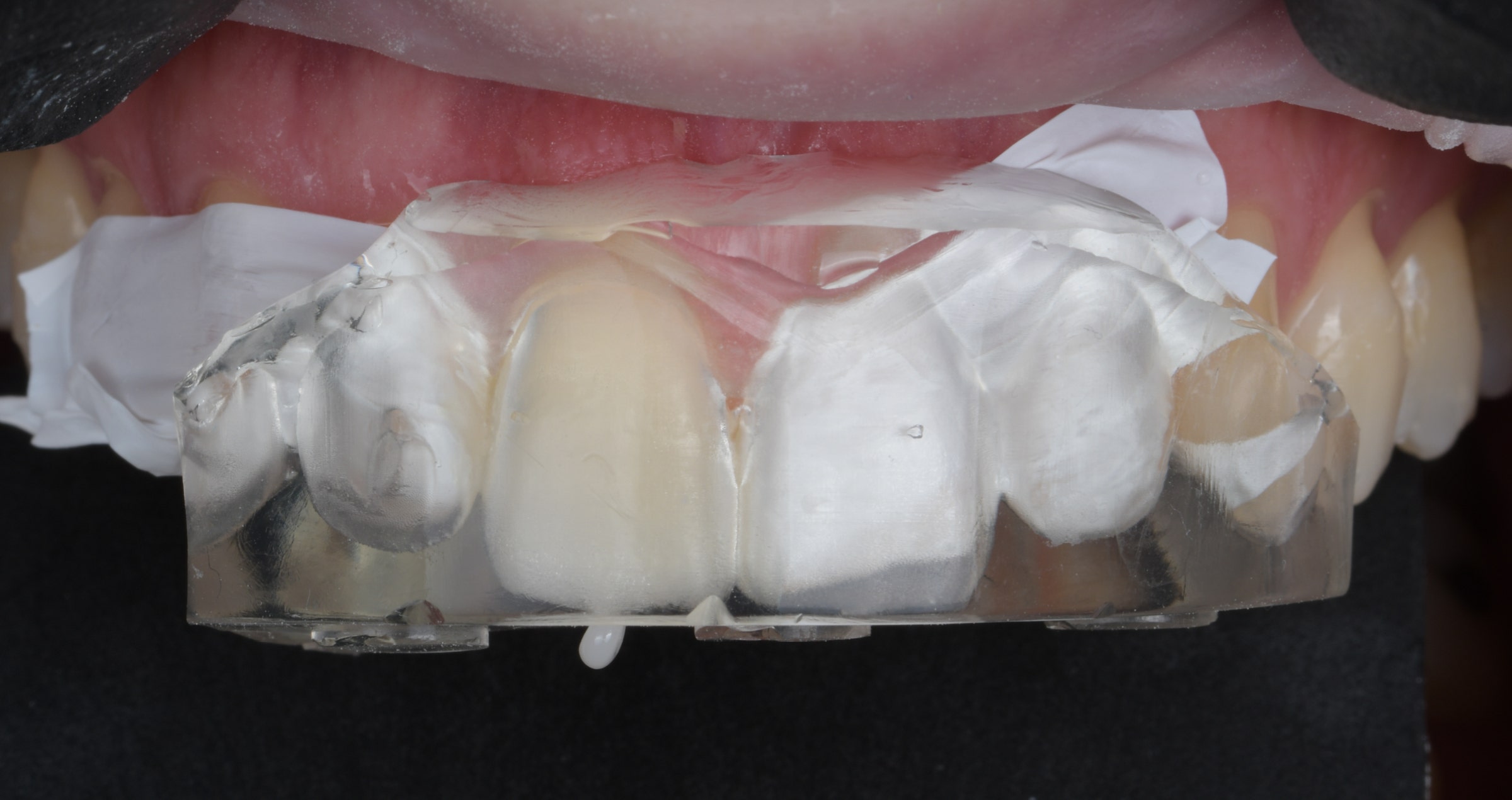
After its application, the silicone index was placed in the mouth and CLEARFIL MAJESTY™ ES Flow Low in the shade A1 injected tooth by tooth through the injection channels (fig. 14). What followed was proper light curing of the composite through the index. The situation after index removal is shown in figure 15. At this stage, the excess still needed to be removed, before protecting and separating the already restored teeth with PTFE tape and repeating the procedure for the other lateral incisor, central incisor and canine.


Fig. 14. Index with injected composite.


Fig. 15. Result after index removal.


Treatment outcome

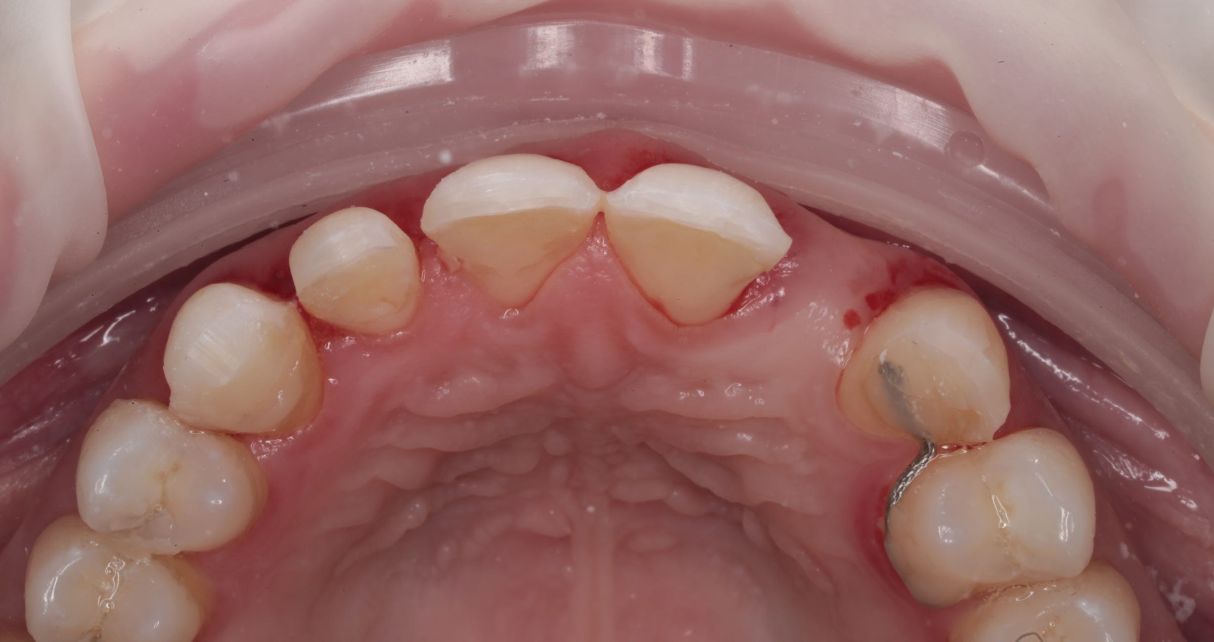
Once all the excess material was completely removed, the occlusion was checked and adjusted. Finishing and polishing was accomplished with TWIST™ DIA for Composite (Kuraray Noritake Dental Inc.). The immediate treatment outcome is shown in figures 16 to 18, while figures 19 and 20 were taken at a recall after six months.


Fig. 16. Treatment outcome: Frontal view.


Fig. 17. The new smile.


Fig. 18. Treatment outcome: Occlusal view.


Fig. 19. Appearance of the teeth…


Fig. 20. … at the six-month recall.


Conclusion

Thanks to advances in restorative materials and technological tools, it is nowadays possible to restore our patients' smiles quickly and reproducibly. The flowable injection technique is one of the most successful examples of modern aesthetic treatments using composite resin. CLEARFIL MAJESTY™ ES Flow has all the qualities needed to carry out these treatments under the right conditions.


About the Author

Dr. Adrien Lavenant obtained his degree in Dentistry from Aix-Marseille University in 2010. He pursued post-university training in periodontology, implantology, and restorative and aesthetic dentistry (Aix-Marseille University and Paris). Dr. Lavenant has been a former teaching staff member at Aix-Marseille University since 2011 and continues to teach in the postgraduate programme in restorative and aesthetic dentistry. He practices in his private clinic in Aix-en-Provence, specializing in restorative, prosthetic, and aesthetic implant dentistry. He has been a member of the international Bio-Emulation group since 2019 and shares his patient care philosophy in accordance with the principles of biomimetic dentistry.


Kiyoko Ban - A legacy in the field of dental technology

By Manabu Suzuki, Director of Dental Division, Kuraray America, Inc.


Kiyoko Ban, a prominent figure in the dental technology field, has made a lasting impact as a researcher, developer, and founder of Noritake Dental business (Fig. 1). Renowned for her contributions to dental porcelains like Noritake's CZR and EX-3, and KATANA™ Zirconia, Ms. Ban stands as a pivotal force in the global advancement of dental technology, earning her the esteemed reputation of developer and marketer within the dental technology community.


After completing her university education in Nagoya, Ms. Ban initially assisted in her family's gas station business. However, driven by a desire for a career change, she enrolled in a newly established dental technician college in Nagoya at the age of 30. Her aspiration was to enter a field where gender distinctions held no sway, offering the potential for worldwide recognition based on technical mastery.


In 1977, a college-sponsored tour to American dental laboratories ignited Ms. Ban's dream to work in the United States. However, she delved into research across various fields such as chromatology (the science of color), ceramics and metals, finding a newfound passion for research over clinical work after graduation because she was offered a "Curriculum Chief" position from the college when she graduated (Fig. 2).


Fig. 2. Ms. Ban, a curriculum chief at the Dental Technicians College, devoted her evenings to material research.


Fig. 3. In the 1990s, Ms. Ban actively engaged in promoting EX-3 through sales efforts in Italy.


Her teaching career spanned from the age of 34 to around 40, during which she pioneered porcelain training sessions for technical improvement and arranged lectures over weekends by famous speakers such as Masahiro Kuwata.


At the age of 40, she resigned teaching career and pursued her research career. The opportunity to conduct full-scale experiments led her to the discovery of a company with advanced ceramic technology "Noritake Co., Limited", renowned for its tableware. In 1986, Cusp Dental Supply, a research institute, was established by Ms. Ban in Nagoya, focusing on the development of materials for PFM crowns. The commercialization of Super Porcelain AAA (EX-3) in 1987 marked a significant milestone, addressing issues prevalent in porcelain materials of that time, such as cracks, greening, and fluorescence.


She began traveling all over Japan and around the world to sell the products she had developed and went on to develop new products that were needed by dental technicians worldwide (Fig. 3). She continued to develop new products such as CZR, CZR Press, and KATANA™, the world's first multilayer zirconia.


Ms. Ban has been actively involved in mentoring students and graduates seeking opportunities to work overseas. During summer vacations, she took students and professionals interested in working abroad to countries like Australia, Germany, and the United States. The aim was to visit dental clinics, dental technician schools, and laboratories, fostering exposure and learning in an international context.


Simultaneously, Ms. Ban delved into researching non-precious dental technology. Inspired by her exposure to the term "non-precious" during her time in the United States, she anticipated its potential in Japan. Her research presented at lectures and events highlighted the shift in the landscape as the price of gold surged, rendering precious alloys containing significant amounts of gold impractical for PFM crowns.


As the demand for their developed products grew, the need for global acceptance became apparent. In 1990, Cusp Dental Research was established in Manhattan, New York, marking Ms. Ban's foray into establishing a company overseas. Despite the unfamiliarity with legal procedures and the challenges of setting up a foreign company, Ms. Ban, driven by determination, overcame these hurdles. The establishment of the company in the United States expanded their presence internationally Fig. 4).


Fig. 4. Capturing the essence of ISC 1996 - the International Symposium on Ceramics in Orlando, FL..


Noritake Dental Supply Co., Limited was established in 1998 by the Noritake Co., Limited, which aimed to further expand its dental business. Despite the absence of a capital relationship with Noritake at the time of establishing the research laboratory, Ms. Ban played a key role in joint research efforts with Noritake. Then she was invited to this company as the position of president, owning 60% of the stock, while Noritake held 40% (Fig. 5).


Besides Noritake Dental business, she continued expansion with the establishment of a dental laboratory in Boston in 1995. Despite the challenges posed by the September 11, 2001 World Trade Center incident, they acquired their building in Boston, integrating their New York laboratory into the Boston operations.


Ms. Ban's tenure as president of Noritake Dental Supply persisted until 2009, but organizational changes following the merger with Kuraray in 2011 led to her transition into an advisory role (Fig. 6). Despite the shift in responsibilities, her commitment to the dental technician profession remained steadfast.


Fig. 5. Noritake Dental Supply Inc Inauguration Party, 1998.


Kiyoko Ban's path encapsulates not just a career but a legacy in the field of dental technology. From her early struggles in a tooth carving class to establishing and expanding international laboratories, Ms. Ban's story is one of determination, innovation, and a deep-rooted commitment to advancing the dental technician profession.


Fig. 6. A scene from Ms. Ban’s retirement celebration as Noritake Dental Supply president, surrounded by esteemed dental technicians from around the world.