429 Too Many Requests

429 Too Many Requests


nginx

Blog

Clearfil Majesty™ ES Flow mottar “NIOM-testet” – kvalitetssikring

CLEARFIL MAJESTY™ ES FLOWMOTTAR “NIOM-TESTET” – KVALITETSSIKRING

Før man kan markedsføre en kompositt som et odontologisk restaureringsmateriale, må den også møte ISO-standard 4049:2019 - Odontologiske Polymerbaserte Restorative Materialer. Grunnet den enormt positive feedback Kuraray Noritake Dental Inc. mottok fra brukere av CLEARFIL MAJESTY™ ES Flow – produktene, ba vi NIOM (Nordisk Institutt for Odontologiske Materialer) om å uavhengig teste disse produktene innen den gjeldende ISO-standard.


Selv om det ikke var obligatorisk eller nødvendig for Kuraray Noritake å få CLEARFIL MAJESTY™ ES Flow ytterligere testet, gjorde vår tro på og selvsikkerheten på kvaliteten i vårt eget produkt at vi også ville gjøre dette. NIOM møysommelig evaluerte CLEARFIL MAJESTY™ ES Flow i alle de tre viskositetene: Høy, medium og lav (fig. 1). Testingen og evalueringen som ble gjort inneholdt herdedybde, bøyestyrke, vannopptak og løselighet, og fargestabilitet etter bestråling og vannopptak. NIOM fant at alle tre viskositeter, inkludert i forskjellige farger av Vita-skalaen, møtte ISO-standardene.

Vi er tilfredse med å ha gått den ekstra distansen og stolte over at en uavhengig tredjepart i NIOM bekreftet at vårt produkt møter de strenge ISO-standardene.


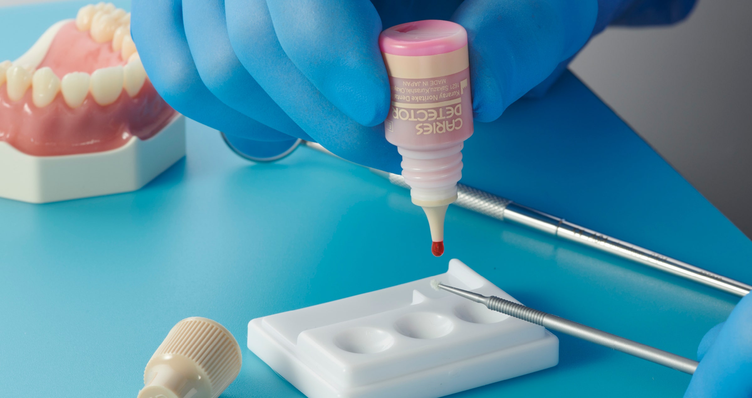
Fig. 1: CLEARFIL MAJESTY™ ES Flow i sine tre forskjellige grader av viskositet, og således flyteegenskaper.


INDIKASJONER FOR KLINISK BRUK

Disse testresultatene er en ekstern bekreftelse for klinikerene at de kan trygt bruke denne populære flytende kompositt-serien slik som spesifisert av Kuraray Noritake Dental Inc, i sine «instructions for use», bruksanvisning. NIOM-testingen hva gjelder herdedybde viser at ved å legge den anbefalte tykkelsen av hvert lag, vil kompositten herde tilstrekkelig – noe som er helt essensielt for langtidsytelsen. I tillegg gir alle tre viskositetene nok mekanisk styrke og egenskaper hva gjelder vannopptak, til å også kunne brukes i restaureringer av okklusalflater posteriort, i premolarer og molarer. Dette betyr at materialene egner seg godt til mange indikasjonsområder, som alle klassifiseringer av fyllinger, samt reparasjoner av kompositt og sementering (Fig. 2).

Fig. 2: De tre varianter av CLEARFIL MAJESTY™ ES Flow og anbefalte indikasjonsområder.


FLOTT ESTETIKK OG GODE HÅNDTERINGSEGENSKAPER

I tillegg til disse gode og velbalanserte mekaniske egenskapene, er CLEARFIL MAJESTY™ ES Flow enkel i bruk grunnet sin innovative appliseringsform fra sprøyte. Dette gjør at man unngår luftblærer, lett kan plassere og anpasse materialet, unngår klebrighet og enkelt kan polere. Materialet dekker nok av fargeskalaen (Fig. 3) og med sin gjennomlysningsevne gir dette en kamelon-effekt slik at det blender pent inn med omliggende tannsubstans, noe som gir et naturlig utseende og pen morfologi. Både estetikk og håndteringsegenskaper har fått gode til svært gode anmeldelser av konsulenter innen dental rådgivning, da gjennom klinisk evaluering.


Fig. 3: Oversikt over fargeskalen per hver enkelt viskositet.


NIOM bekrefter også de gode estetiske egenskapene gjennom instituttets testmetoder for fargestabilitet etter vannopptak og bestråling. Disse viser CLEARFIL MAJESTY™ ES Flow sin stabilitet over tid, noe som er viktig for materialets overlevelsesprognose i munnen og langtidsestetikk.


Velg et pålitelig, høykvalitets, flytende, direkte komposittmateriale som tåler strenge testkrav. Klikk her for mer informasjon om CLEARFIL MAJESTY™ ES Flow.


BEST.FIT: A hybrid technique for an efficient and aesthetic restoration of anterior teeth

Case by Dr. Enzo Attanasio

The introduction of new-generation composites, equipped with nanofillers and highly loaded, has opened doors to new techniques for managing direct and semi-direct restorations. In particular, over the last ten years, there has been a significant revolution in the world of flowable composites. Nowadays, these materials offer a filler percentage very similar to packable composites through precise interventions in resin matrix management. They come in various viscosities, offering numerous advantages both in terms of handling and clinical use, as well as beneficial mechanical and physical characteristics.


FLOWABLE INJECTION TECHNIQUE

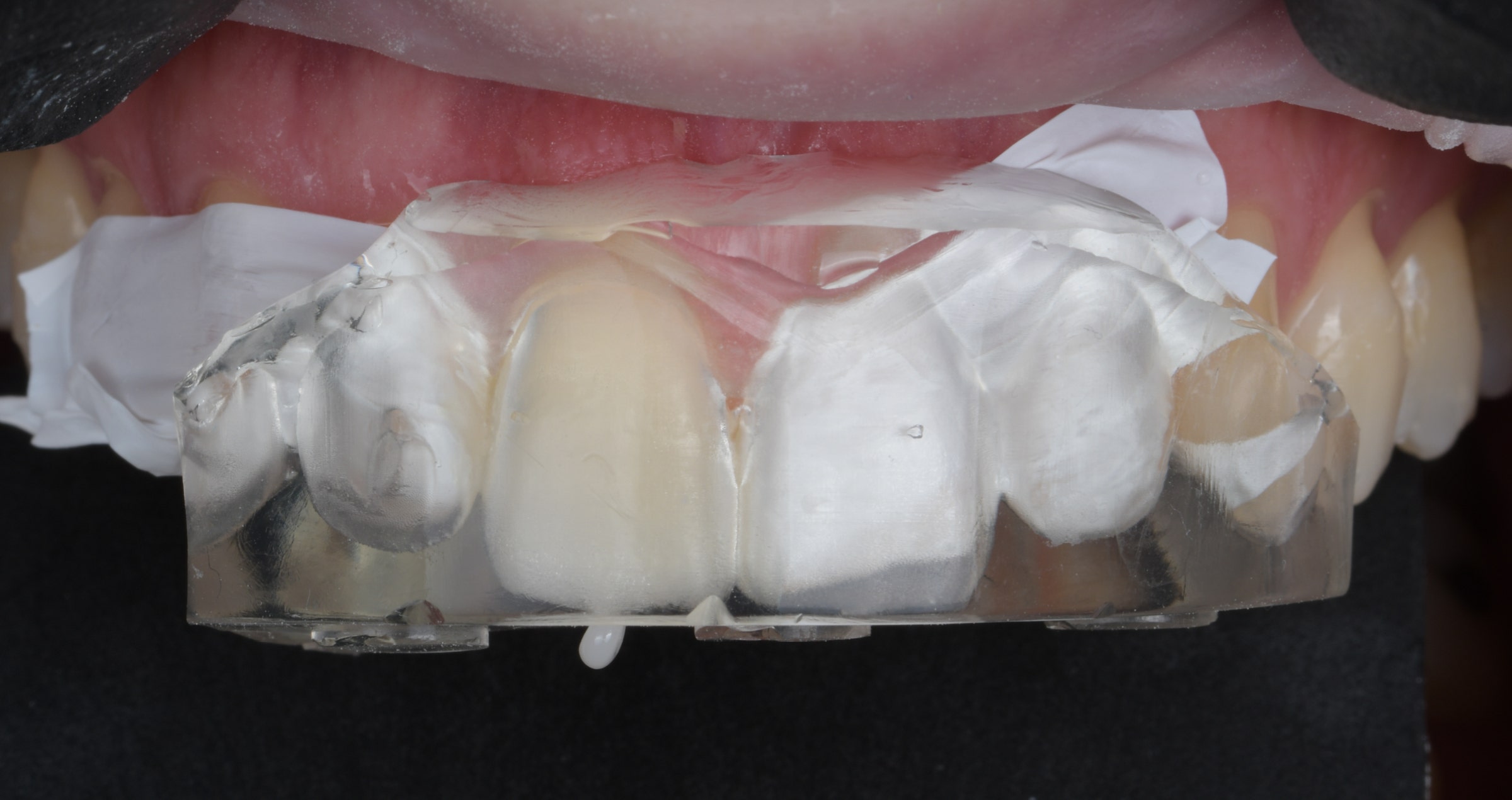
This new era of flowable composites has seen the development of a technique known as the Flowable Injection Technique (also referred to as injection moulding). It enables dental practitioners to reproduce anatomical forms created by a dental technician in the laboratory through a diagnostic wax-up. The shapes planned on the model are transferred directly in the patient's mouth using transparent silicone matrices or indexes, into which the composite is injected through specific injection holes.


The main difference compared to traditional mock-ups is that the reproduced dental elements remain separate from each other. This technique provides predictable results identical to those developed on the technician's wax-up, requiring less chair time than direct veneering and offering a longevity similar to traditional composite restorations.


BENEFITS AND CHALLENGES

The major benefit of this technique is the faithful reproduction of morphological details that the technician creates on the diagnostic wax-up, which the clinician can reproduce with minimal effort. The restoration produced through the flowable injection technique, if all steps are followed correctly, requires minimal finishing by the clinican, who only needs to focus on polishing the composite.


However, one limitation is the difficulty in isolating the operative field, often requiring a split-dam technique or labial retractors, with all the associated adhesive challenges. The use of a rubber dam is only feasible if the peripheral dental tissues around the restoration are euchromatic, allowing the technician to create a wax-up with supragingival preparation margins.


Another compromise with the flowable injection technique is the management of the composite as a single mass. This makes it only possible to reproduce natural incisal translucencies typical of young patients by performing complex cutbacks and subsequent incisal painting. Without specific operator skills, the outcomes of this time-consuming manual procedure are unpredictable.


HYBRID TECHNIQUE: BEST.FIT

To leverage the advantages of both classical direct anterior restoration and flowable injection techniques and eliminate the limitations, a hybrid technique known as BEST.FIT (Buccal Enamel Shade Through Flow Injection Technique) has emerged. This technique allows the operator to manage the delicate phase of reproducing the buccal enamel layer of the anterior restoration through the flowable injection technique, keeping certain aspects in mind during the injection phase.


PROCEDURE

The transparent silicone key used for the creation of the buccal enamel layer is similar to the one used in the original flowable injection technique. The initial phase of restoration management follows all the classical steps of direct technique, requiring isolation with rubber dam. The palatal enamel layer is recreated with a highly translucent packable composite, and the palatal portion of the interproximal walls is produced using a suitable matrix system. Then, the core of the restoration is defined with opaque masses, creating mamelons and adding incisal effects. It's crucial to control the residual enamel thickness using a vestibular silicone index, aiming for about 0.3 mm of space. The buccal portion is finally reconstructed during the injection phase. The transparent silicone index created on the wax-up should be tested after each reconstruction phase to ensure passive insertion.


After creating the restoration core, the element to be injected is separated from the contiguous ones with thin PTFE tape. The transparent mask is then inserted, and fluid composite is injected through the injection holes to precisely reconstruct the buccal enamel thickness. The composite tip should be positioned at least halfway through the buccal surface, and the injection should be slow and controlled to avoid air bubbles in the material.


FINISHING

Following a 40-second polymerization vestibularly and occlusally, the transparent matrix is carefully removed, and excess interproximal composite above the PTFE tape as well as any remaining composite cylinder from the injection holes are removed. After completing all restorative elements, the rubber dam is dismantled, and composite excess is finished. After checking the occlusion, the composite is polished, usually requiring no further intervention.


CASE EXAMPLE

Fig. 1. Female patient with discoloured anterior restorations desiring a smile makeover.


Fig. 2. Close-up of her maxillary anterior teeth.


Fig. 3. Restorations in need of replacement: Lateral view from the right.


Fig. 4. Restorations in need of replacement: Lateral view from the left.


Fig. 5. Printed model based on a digital diagnostic wax-up based on a digital impression.


Fig. 6. Palatal silicone index produced for the conventional direct restoration steps.


Fig. 7. Transparent matrix with injection holes produced for the build-up of the buccal enamel layer using the flowable injection technique.


Fig. 8. Operative field isolated with rubber dam.


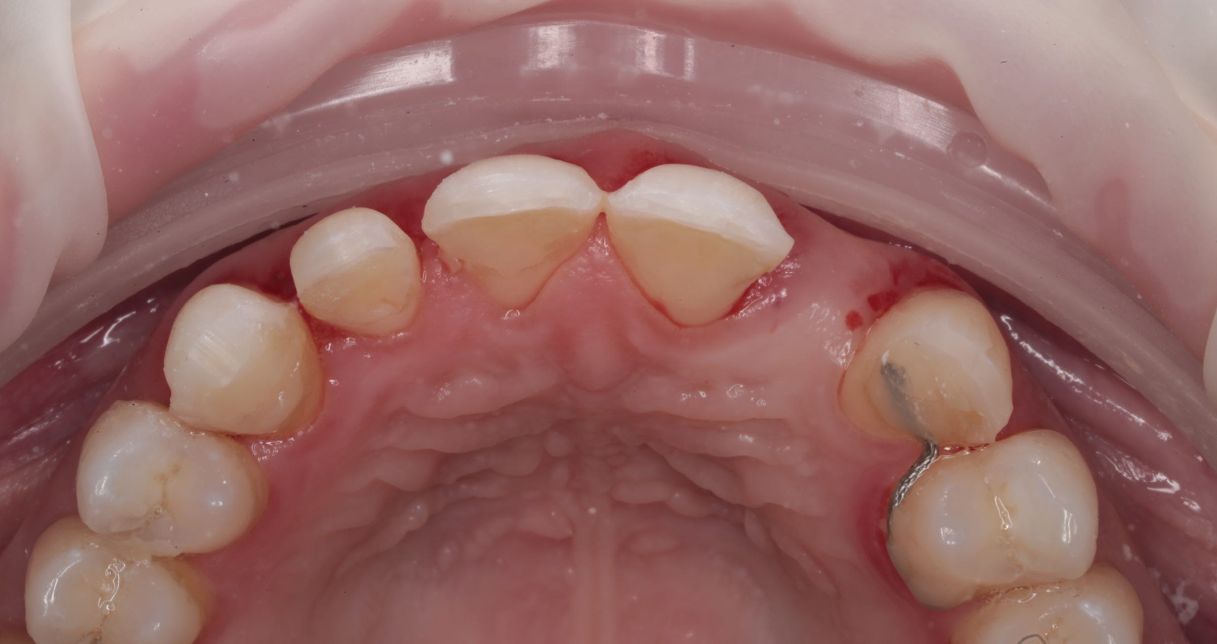
Fig. 9. Existing restorations removed and tooth surfaces roughened at the start of treatment.


Fig. 10. Palatal silicone index positioned intraorally for the build-up of the palatal wall.


Fig. 11. Checking of the space available in the vestibular area with a second silicone index.


Fig. 12. Etching with phosphoric acid etchant.


Fig. 13. Application of a universal adhesive (CLEARFIL™ Universal Bond Quick, Kuraray Noritake Dental Inc.).


Fig. 14. Palatal walls built up with CLEARFIL MAJESTY™ ES-2 Premium in the shade A1E with the aid of the palatal silicone index.


Fig. 15. Build-up of the interproximal walls with CLEARFIL MAJESTY™ ES-2 Premium in the shade A1D and establishing of the contact points using anatomical sectional matrices for the posterior area placed vertically.


Fig. 16. Dentin core built up with CLEARFIL MAJESTY™ ES-2 Premium in the shade A2D. CLEARFIL MAJESTY™ ES Flow Super Low in the shade XW was applied on the mamelons, while CHROMA ZONE™ COLOR STAIN Blue (Kuraray Noritake Dental Inc.) was used to reproduce incisal translucencies in the spaces not covered by the dentin core.


Fig. 17. Try-in of the transparent matrix for flowable injection.


Fig. 18. Isolation of the adjacent teeth with PTFE tape for a one-by-one injection.


Fig. 19. CLEARFIL MAJESTY™ ES FLOW Low in the shade A2 (Kuraray Noritake Dental Inc.) injected for the anatomical shaping of the maxillary right central incisor.


Fig. 20. Situation after flowable injection for all four anterior teeth, light curing through the matrix, final matrix removal and excess removal.


Fig. 21. Treatment outcome …


Fig. 22. … with visible mamelons, natural incisal translucencies …


Fig. 23. … and a lifelike anatomical shape …


Fig. 24. … of the restorations.


CONCLUSION

Each work phase must be executed with extreme care to lay the foundations for a passive linking of all subsequent steps without creating difficult management situations. The BEST.FIT technique is a convenient and useful method for dental practitioners to manage multiple direct anterior restorations simply and predictably, especially in situations requiring complex rehabilitations with large restorations.


Dentist:

ENZO ATTANASIO


Enzo Attanasio graduated in 2008 in Dentistry and Dental Prosthetics from the Magna Graecia University of Catanzaro. In 2009, he went on to specialize in the use of laser and new technologies in the treatment of oral and perioral tissues at the University of Florence. That year he also attended Prof. Arnaldo Castellucci’s course in Clinical Endodontics at the Teaching Center of Microendodontics in Florence where, in 2012, he went on to complete his training in Surgical Microendodontics. In 2017 he attended a course on Direct and indirect Adhesive Restorations at Prof. Riccardo Becciani’s Think Adhesive training center in Florence where he later become a tutor. Today, as a member of the Italian AIC and based in Lamezia Terme, Italy, Dr Attanasio has a special interest in Endodontics and Aesthetic Conservative.


Article by Dr. Michał Jaczewski

FLOWABLE INJECTION AND STAMP TECHNIQUE: RESTORING TEETH IN THE POSTERIOR REGION

Restoring the occlusal surface of posterior teeth while preserving the natural morphology and re-establishing correct occlusal contacts has always been challenging for dental practitioners. Free-hand layering requires knowledge of tooth anatomy, composite handling skills and experience. When the occlusal surface of a tooth is damaged at the start of treatment (as is usually the case in teeth with large MOD cavities) or an increase of the vertical dimension of occlusion is planned (e.g. in severely worn teeth), the use of the flowable injection technique may be a suitable alternative. It truly speeds up and facilitates the process of building up the restoration to a natural shape, but requires thorough planning and preparation. In cases with an intact occlusal surface, the stamp technique might be the first choice.


FLOWABLE INJECTION TECHNIQUE: GENERAL CONSIDERATIONS

It is up to the user how exactly the restorations, to be built up by flowable injection, are planned and how the plan is implemented: One can either opt for a conventional wax-up or make use of digital tools in the planning phase. Dedicated design software offers the benefit of facilitating the creation of a natural shape and morphology of the desired restoration and allows for the establishing of an ideal occlusal relationship. Once the wax-up is ready, it needs to be transferred into the patient’s mouth. This is accomplished via a printed or classical model with wax-up, which forms the basis for the production of a matrix or silicon index. This index is then used intraorally for the injection of the flowable composite. To enable proper light curing through the index, the index material should be as transparent as possible.


AREA-SPECIFIC CONSIDERATIONS

In the posterior area, an index made of two different materials – a soft inner silicon structure and a hard outer shell – may be advisable. Due to its higher dimensional stability compared to a soft silicon index, it is possible to put pressure on it for proper adaptation to the isolated teeth and soft tissue without the risk of altering the shape of the tooth. Figure 1 shows such an index on and next to a printed model. It consists of a hard shell made of acrylic and a soft inner structure made of a transparent silicone material (e.g. EXACLEAR™, GC). For production, a high-capacity hydraulic pressure curing unit designed for use with self-curing resins (Aquapres™, Lang Dental) has proven its worth: It ensures a highly accurate reproduction of the (digital) wax-up.


Fig. 1. Printed model and silicone index.


Reconstruction of posterior teeth with the flowable injection technique requires prior removal of all carious lesions and reconstruction of the proximal surfaces to restore the contact points. Hence, the injected composite serves the exclusive purpose of restoring the occlusal surface. When several teeth are treated, a two-step procedure with an alternating technique is recommended to provide for proper separation of the teeth. Blocking the proximal surfaces below the contact point with PTFE tape will reduce the amount of excess material in these areas and make it easier to clean and prepare the proximal surfaces after flowable injection. Proximal and deeper occlusal lesions should be restored with the aid of a matrix, wedge and ring.


CLINICAL PROTOCOL

A possible clinical protocol is illustrated in Figures 2 to 5: After caries excavation and tooth preparation, sectional matrices, wedges and rings were placed to allow for simultaneous treatment of the mesial and occlusal cavities. Following etching and application of the universal adhesive CLEARFIL™ Universal Bond Quick (Kuraray Noritake Dental Inc.), the cavities were restored with CLEARFIL MAJESTY™ ES Flow Super Low in the shade A1 and CLEARFIL MAJESTY™ ES-2 Universal in the shade U. The distal cavity of the first molar was filled in the last step of the free-hand modeling procedure. In order to restore the occlusal surfaces in their original vertical dimension, every second tooth was isolated with rubber dam and the exposed molar etched (total-etch technique with K-ETCHANT Syringe, Kuraray Noritake Dental Inc.). the alternating index was positioned with some pressure and the flowable composite (CLEARFIL MAJESTY™ ES Flow Super Low) injected. Once light curing was completed, it was possible to remove the index, chip off the excess and finish and polish the restoration before repeating the procedure for the adjacent molar.


Fig. 2. Restoration of two molars: Teeth preparation and caries excavation.


Fig. 3. Restoration of two molars: Filling of the proximal and occlusal cavities.


Fig. 4.  Restoration of two molars: Re-establishing the occlusion with the aid of the flowable injection technique.


Fig. 5. Alternating approach: Restoration of the second molar by injecting flowable composite.


DISCUSSION

The use of the flowable injection technique allows for rapid restoration of teeth and the establishment of precise occlusal contacts. This reduces the time spend on occlusal surface modelling and minimizes the risk for prolonged treatment due to a repeated need for occlusal adjustments. In addition to saving time, it is possible with this technique to restore a greater number of teeth in a single appointment. The aesthetics of this type of restoration may be somewhat limited: A skilled practitioner is able to achieve better aesthetic results on the occlusal surface. However, with a detailed wax-up and high-quality model great outcomes can be obtained. The surface quality of printed models can be increased by adjusting the printing parameters including the layer height (Fig. 6). The use of a hydraulic pressure curing unit for silicone index production further increases the quality of the occlusal surface.


When planned and implemented correctly, the established occlusal surface and contacts reflect the natural anatomy without the need for adjustments (Fig. 7). Especially when restoring an entire quadrant, it is possible to increase the efficiency by opting for the flowable injection technique. Doing so reduces the number of appointments and the chair time decisively (Fig. 8).


STAMP TECHNIQUE: CONSIDERATIONS

If the occlusal surface of the tooth is intact, a wax-up may not be necessary. In this case, the better strategy is to duplicate what is still available before initiating treatment. A flowable composite or liquid rubber dam can be used for this purpose. It is important to coat the tooth surface with glycerin gel before applying the material. This will facilitate separation of the stamp from the tooth. It is always advisable to create a stamp that covers not only the details that need to be recorded and duplicated, but is extended over the cusps. This offers better stability in the restoration phase.


CLINICAL PROTOCOL

Figures 9 to 11 illustrate a possible clinical procedure. In this case, a molar with an occlusal carious lesion needed to be restored. The tooth surface was cleaned and a thin layer of glycerin gel applied, followed by a thick layer of liquid rubber dam, which covered the entire occlusal surface. Then, a micro applicator was immersed into the material and the stamp cured. After preparation, etching and application of the bonding system, the cavity was restored with flowable composite (CLEARFIL MAJESTY™ ES Flow Super Low in the shade A2). When the cavity is larger and depending on personal preferences, a paste-type composite (CLEARFIL MAJESTY™ ES-2 Universal) may also be used. Prior to light curing of the composite, the occlusal surface was covered with PTFE tape and the stamp pressed onto it. After firm pressing, the tape and excess material were removed and the restoration polymerized. This restoration faithfully reproduces the occlusal surface and did not require any occlusal adjustments.


Fig. 6. Stamp production with liquid rubber dam.


Fig. 7. The stamp.


Fig. 8. Restoration procedure: From preparation to bonding.


Fig. 9. Restoration procedure: Filling with flowable composite.


Fig. 10. Restoration procedure: Duplication the original occlusal surface with the stamp.


Fig. 11. Tooth before and after treatment using the stamp technique.


CONCLUSION

Techniques that add simplicity and efficiency to clinical procedures are always welcome in the busy practice environment. Depending on the information available at the start of treatment and the number of teeth to be restored, the flowable injection or the stamp technique may be an ideal choice. They are easily implemented and speed up the clinical procedure, but most importantly support predictable outcomes. This saves time in the finishing phase and minimized the risk of repeated adjustments, hence protecting everyone involved from additional appointments and frustration. Especially for practitioners with limited routine in free-hand modelling and for those with maximum patient comfort in mind, both techniques are worth being integrated in their clinical procedures.


Dentist:

MICHAL JACZEWSKI


Michał Jaczewski graduated from Wroclaw Medical University in 2006 and today runs his private practice in the city of Legnica, Poland. He specializes in minimally invasive dentistry and digital dentistry and is the founder of the Biofunctional School of Occlusion. Here he lectures and runs workshops with focus on full comprehensive patient treatments.


Universal: Whenever a high translucency is desired

Case by Dr. Jusuf Lukarcanin


In teeth in which the areas to be restored are surrounded by a lot of non-discoloured tooth structure - as may be the case in Class I, II and Class V cavities - the use of CLEARFIL MAJESTY™ ES-2 Universal in the shade U may be an option. The 28-year-old patient, who presented for diastema closure, had teeth with a comparatively low translucency and different shades due to smoking and excessive coffee consumption. As the composite was applied in enamel areas only, the relatively high translucency of the universal shade seemed beneficial in this case.


Fig. 1. Initial clinical situation.


Fig. 2. New smile of the patient.


Reasons for selecting universal:

- Large amounts of underlying or surrounding tooth structure present

- Medium light-scattering desired


Universal properties:

- High translucency

- Medium light-scattering effect


Dentist:

JUSUF LUKARCANIN


Dr. Jusuf Lukarcanin is a Certified Dental Technician (DCT) and a Doctor of Dental Science (DDS). He studied dentistry at the Ege University Dental Faculty in Izmir, Turkey, where he obtained a Master‘s degree in 2011. In 2017, he received a Ph.D. degree from the Department of Restorative Dentistry of the same university. Between 2012 and 2019, Dr. Lukarcanin was the head doctor and general manager at a private clinic in Izmir.


Between 2019 and 2020, he worked at Tinaztepe GALEN Hospital as a Restorative Dentistry specialist, between 2020-2022 he worked at MEDICANA International Hospital Izmir as a Restorative Dentistry specialist. Currently he is an owner of a private clinic for aesthetics and cosmetics in Izmir.


Restoration of a single central incisor: Mastering the art of observation

Case by Andreas Chatzimpatzakis


Observe and copy: This is the key to nature-like dental restorations. There are many optical effects, colour transitions and morphological details in natural teeth that need to be taken in and understood – and replicating them is only possible for those who know exactly how their materials work. Once these skills are acquired, however, they enable a dental technician to produce their restorations as truly beautiful copies of nature. Even when restoring a single maxillary central incisor, the technique delivers outstanding – or inconspicuous - outcomes, as revealed by the following example.


Using high-quality, translucent and gradient-shaded zirconia frameworks and porcelains, the layering technique does not have to be highly complicated. Two bakes and a number of selected effect liquids, internal stains and porcelains are usually sufficient for outcomes that exceed expectations.


CASE EXAMPLE


In the present case, a young male patient had a quite opaque crown on his maxillary right central incisor that needed to be replaced. During shade selection in the dental laboratory (Fig. 1), it was observed that the cervical third of the adjacent central incisor is lighter than the rest. Its shade in other areas corresponded to B4 on the VITA classical A1-D4® Shade Guide. Hence, it was decided to use a somewhat lighter material for the framework and darken the restoration especially in the middle and incisal areas with internal stains.


The concrete plan was to mill a coping made of KATANA™ Zirconia STML (Kuraray Noritake Dental Inc.) in the shade A3, characterize it with Esthetic Colorant (both Kuraray Noritake Dental Inc.) and sinter the piece (Figs 2 to 4). In the following layering procedure including just two bakes, a combination of internal stains and selected porcelains (CERABIEN™ ZR, Kuraray Noritake Dental Inc.) was applied as illustrated in Figures 5 to 12. Figures 13 to 17 display the result on the model, minor adjustments during try-in and the final treatment outcome.


Fig. 1. Shade selection. The cervical third of the adjacent central incisor is lighter than usual compared to the middle and incisal areas.


Fig. 2. Coping made of KATANA™ Zirconia STML in the shade A3.


Fig. 3. Intensification of some shade characteristics of the multi-layered blank using Esthetic Colorant in the shades Grey (middle) and Blue and Grey (incisal area).


Fig. 4. Coping after sintering.


Fig. 5. Colour map for internal staining, using CERABIEN™ ZR Internal Stains.


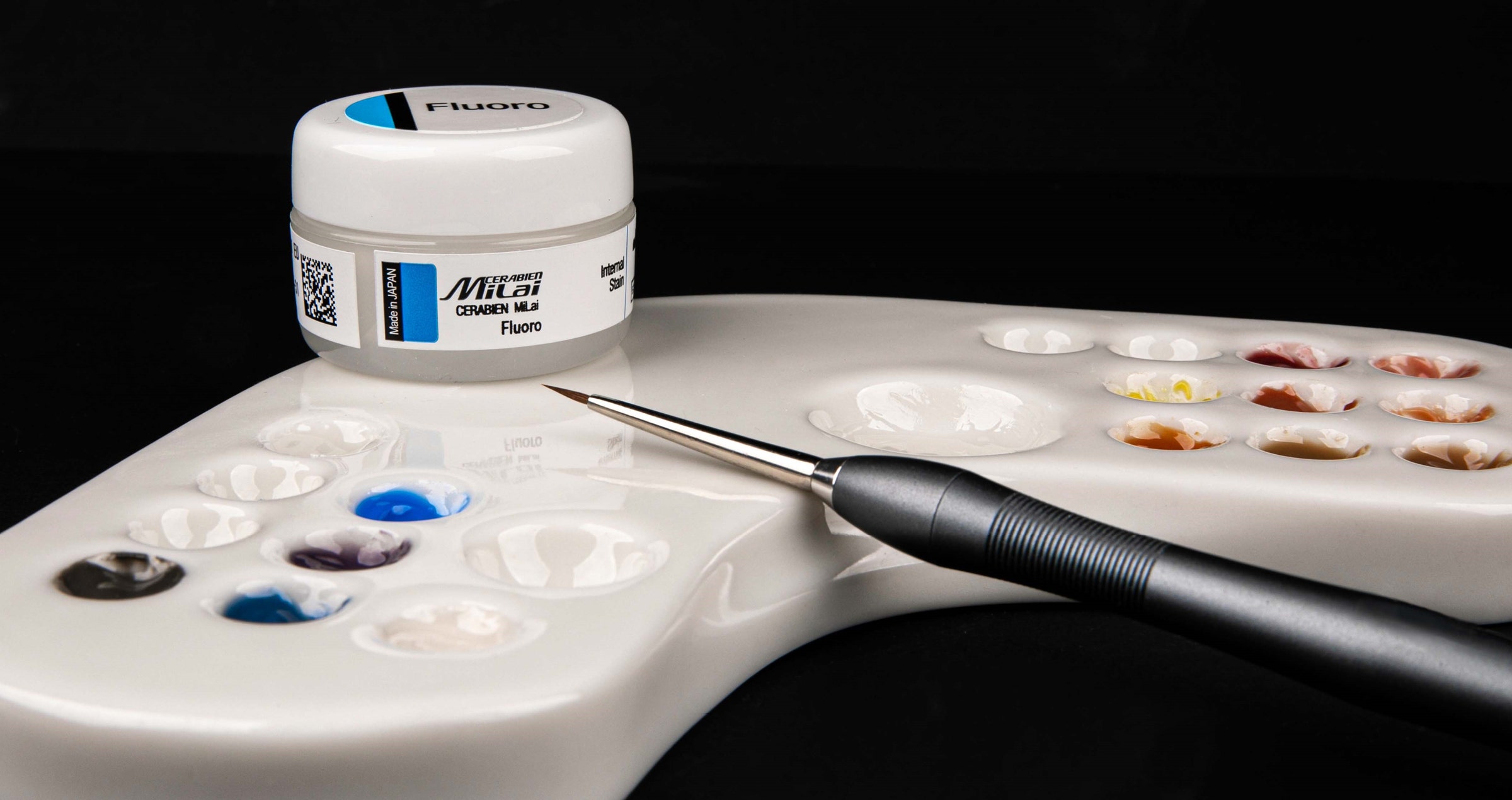
Fig. 6. Result of the use of Shade Base Stain Modifier Fluoro to increase the fluorescence and internal staining as planned.


Fig. 7. Application of Opacious Body OBA2, …


Fig. 8. … Translucent Tx …


Fig. 9.: … and Luster CCV-2.


Fig. 10. Crown after the first bake.


Fig. 11. Crown after the application of CERABIEN™ ZR Internal Stains: A+, Aqua Blue 2, White mixed with Cervical 2 (ratio: 30/70) for the cracks, and Cervical 2.


Fig. 12. Application of Luster LT1 to finalize the shape.


Fig. 13. Finished crown after the second bake on the model.


Fig. 14. Evaluation of the surface texture: Observing and copying the surface details is as important as the imitation of the shade characteristics.


Fig. 15. Minor texture adjustments during try-in.


Fig. 16. Final restoration in place after cementation with PANAVIA™ V5 (Kuraray Noritake Dental Inc.).


Fig. 17. Treatment outcome.


CONCLUSION


Mastering the art of observing natural teeth is the key to lifelike restorations. It allows a dental technician to develop a deep understanding of shade and morphology, which is – apart from knowing the selected materials very well – the only talent needed to reach a high level of excellence. Those who are observant and take in every detail with their eyes can be sure that their mind will understand and their hands will automatically follow.


Dental technician:

ANDREAS CHATZIMPATZAKIS


Andreas graduated from the Dental Technology Institute (TEI) of Athens in 1999. During his studies he followed a program at the Helsinki Polytechnic Department of Dental Technique, where he trained on implant superstructures and all ceramic prosthetic restorations. As of 2000, he is running the ACH Dental Laboratory in Athens, Greece, specialized on refractory veneers, zirconia and long span implant prosthesis. In 2017 Andreas visited Japan where he trained under the guidance of Hitoshi Aoshima, Naoto Yuasa and Kazunabu Yamanda and become International Trainer for Kuraray Noritake Dental Inc..


Large cavity restoration with resin composite: which materials to choose?

Case by Vasiliki Tsertsidou


What kind of resin composite is recommended for core build-up procedures? While there are specific dual-cure core build-up resin composites available on the market, it is not mandatory to use them. Light curing is advisable to be applied even for materials with dual-cure polymerization. Some conventional resin composites demonstrate more favourable properties for a core build-up compared to specific core build-up resin composites itself.1 Hence, it is possible to utilize a composite generally used in the dental office, provided it is indicated to and it is not applied deep within the root canal, where proper light curing would be impossible. The critical material properties for core build-ups are high filler load, sufficient flexural modulus and flexural strength.


CLEARFIL MAJESTY™ ES-2 composite series (Kuraray Noritake Dental Inc.) are suitable option for this case. With a filler load weight percentage of 78 and a flexural strength of 118 MPa (according to manufacturer), CLEARFIL MAJESTY™ ES-2 Classic corresponds to core build-up prerequisites*. The following case is illustrating the clinical procedure.


*The indication range of CLEARFIL MAJESTY™ ES-2 composite does not cover core build-up. In the specific case it is used for creating a large Class II filling where all conditions from the IFU, such as curing depth, are met.


Fig. 1. Endodontically treated tooth with a vertical fracture of palatal wall on maxillary right second premolar.


Fig. 2. Buccal view of the tooth.


Fig. 3. Clinical image, directly after removal of fragment.


Fig. 4. Fragment of the maxillary right second premolar.


Fig. 5. Circumferential matrix band for build-up to assist endodontic retreatment.


Fig. 6. Build-up of the missing walls (margin relocation) with CLEARFIL MAJESTY™ ES-2 Classic (A3).


Fig. 7. Temporary filling of the cavity.


Fig. 8. Replacement of the temporary filling material with CLEARFIL MAJESTY™ ES-2 Classic.


Fig. 9. Crown preparation.


Fig. 10. Proximal carious lesion present on the adjacent fist premolar.


Fig. 11. Situation after rubber dam placement and caries removal.


Fig. 12. Cavity restored with CLEARFIL MAJESTY™ ES-2 Classic.


Fig. 13. Prepared crown.


Fig. 14. Crown after sandblasting of the intaglio.


Fig. 15. Mechanically cleaned abutment tooth ready for pre-treatment.


Fig. 16. Intaglio of the crown treated with CLEARFIL™ CERAMIC PRIMER PLUS.


Fig. 17. Etching of the composite surface with phosphoric acid gel.


Fig. 18. Air-drying of PANAVIA™ V5 Tooth Primer on the abutment tooth.


Fig. 19. Crown in place after cementation with PANAVIA™ V5 Paste and excess removal.


A GOOD CHOICE


Dual-cure core build-up resin composites are two-component materials that need to be mixed homogeneously, which obstracts composition from containing high filler load. However, to prevent deformation of the core, a highly filled composite is advisable. This better simulates the flexural modulus of natural tissues compared to materials with low filler load. Consequently, a light-curing material like CLEARFIL MAJESTY™ ES-2 might be a better option. Applied in 2-mm increments in the core area (and not in the root canal), it performs well and provides the desired outcomes. Additionaly, the option of utilising the same material as for any other type of direct restorations is simplifying the stock management and supporting dental practitioners striving for a simplification of clinical procedures.


References

1. Spinhayer L, Bui ATB, Leprince JG, Hardy CMF. Core build-up resin composites: an in-vitro comparative study. Biomater Investig Dent. 2020 Nov 3;7(1):159-166. doi: 10.1080/26415275.2020.1838283. PMID: 33210097; PMCID: PMC7646551.


Dentist:

VASILIKI TSERTSIDOU


Anterior crowns on teeth and an implant

Case by Martin Laurik, MDT


There are so many different restorative materials out there and so many design and finishing concepts available that it often seems difficult to select the best option for a specific case. Using an allrounder like KATANA™ Zirconia YML can facilitate decision making: It is a great choice for single- to multi-unit restorations, works on teeth and implants alike, and can be adapted to individual needs by selecting a suitable design concept and adequate finishing technique. In this way, it is even possible to solve aesthetically challenging cases as the one illustrated below.


Initial situation and temporization


This patient was in need for treatment after the loss of her maxillary right central incisor and the placement of an implant in this region. As a replacement of the restorations on the other three maxillary incisors was necessary as well, it was decided to produce four crowns made of the same material – KATANA™ Zirconia YML. For aesthetic evaluation of the restorations’ length, angulations and shape in the mouth and a functional test drive, the crowns were digitally designed in full contour and milled from PMMA in the determined tooth shade A2 (Fig. 1).


Fig. 1. Full-contour PMMA crowns on the master cast.


Design, milling and effect dyeing of the zirconia crowns


Once the appearance and functional aspects of the temporary restorations were approved by the patient and the restorative team, the definitive crowns were produced. Their design was based on the full-contour design of the temporaries; however, a facial reduction of 0.6 mm was carried out by the software to create space for individualization with a small layer of veneering porcelain. The crowns were then milled from a KATANA™ Zirconia YML disc in the shade A1 – approximately one shade lighter than the determined tooth shade. To mask the uneven colour from the tooth stumps and the implant abutment, the intaglio of the crowns was treated with Esthetic Colorant in the shade Opaque. Some individual and intensified colour effects on the vestibular surface were also created with Esthetic Colorant.


Internal staining and porcelain layering


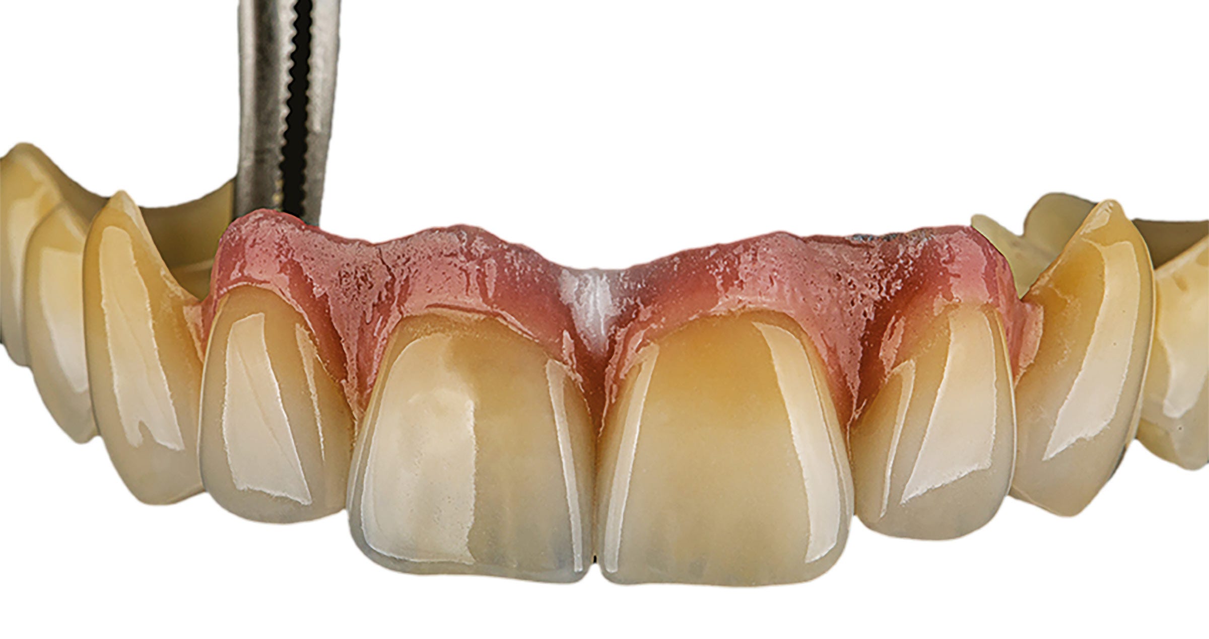
To slightly adjust the chroma and lightness, a first layer of CERABIEN™ ZR Internal Stains was added, followed by a wash bake. After the application of a first layer of CERABIEN™ ZR porcelains (Body, Enamel and Translucent) and baking (Fig. 2) – the central incisors received a layer of A1B, the lateral incisors a mixture of A1B and A2B (slightly darker to provide for a better match with the canines) with LT1, LT Natural completing the picture – additional internal staining was carried out (Fig. 3). The final layer of CERABIEN™ ZR luster porcelains (LT1, ELT2 used on the convex line angles to achieve an external reflection) was added and fixed in a fourth bake (Fig. 4).


After adjustments and very rough polishing, a self-glaze firing programme was selected (firing temperature 915 °C, holding time 5 seconds). On the highly polished incisal and palatal parts of the crowns and for contact point adjustment, CERABIEN™ ZR FC Paste Stain Glaze was applied and fixed with the same bake. The finished crowns on the model are shown in Figure 5, while Figure 6 displays the final treatment outcome.


Fig. 2. Crowns milled from KATANA™ Zirconia YML with a facial cutback of 0.6 mm after individualization with Esthetic Colorant, sintering, internal staining and the application of a first layer of porcelain.


Fig. 3. This picture shows the subtle internal stain adjustment to the ceramic mostly on the incisal part.


Fig. 4. Crowns prior to final shape adjustments and polishing.


Fig. 5. Finished crowns on the model.


FINAL SITUATION


Fig. 6. Treatment outcome.


Easy approach to beautiful restorations


The presented approach is a relatively easy way of producing highly aesthetic anterior restorations. Using an allrounder zirconia combined with a few selected effect liquids, internal stains and luster porcelains, it is possible to achieve a great optical integration even in a situation where teeth and implants need to be restored. The natural shape and surface texture of the restorations plays an important role in this context, as does the base material – a naturally shaded, highly translucent zirconia.


Dentist:

MARTIN LAURIK, MDT


Martin started working as a dental technician in 2014. In the time since, he never stopped training and learning from renowned colleagues. Continuing education courses focused on dental ceramics and occlusion in the functional concept of Slavicek. Fascinated by the beauty of natural teeth, developing an understanding of their complexity and learning how to mimic nature’s design as closely as possible has always been his primary goal, while he is well aware that there is still a lot to be learned and explored on the road to excellence.


A GUIDE TO SUCCESSFUL ZIRCONIA BONDING


Unlock the power of zirconia: perfect for adhesive cementation, the ideal material for a wide range of indications, and essential in minimal invasive dentistry. Time to trust zirconia bonding!

This article demystifies zirconia bonding, providing clear, practical steps to ensure long-term functionality and patient satisfaction, all based on scientific research. Master the three adhesion pillars: mechanical retention, chemical activation, and wetting capacity. Discover how to successfully prepare zirconia surfaces, avoid pitfalls like misapplying silica coating and silane, and choose proven bonding systems for optimal results. Optimise retention even with minimal tooth preparation and achieve reliable zirconia restorations. Say goodbye to doubts and hello to successful zirconia bonding!


Factors influencing retention

Loss of retention due to de-cementation or debonding is a common cause of dental prostheses' failure.  First, let’s have a look at how to cope with the three main factors significantly influencing retention: tooth preparation, restoration pre-treatment, and cement type/bonding.

Tooth preparation

The abutment tooth's height, angle, and surface texture must be considered to achieve sufficient retention and resistance from the preparation. The retention form counteracts tensile stresses, whereas the resistance counteracts shear stresses 4. With the proper preparation, a restoration resists dislodgement and subsequent loss.

Full coverage restorations

To achieve sufficient retention and resistance for full-coverage crowns, the tooth abutment should be at least 4 mm high, and the convergence angle should range from 6 to 12 degrees with a maximum of 15 degrees 1, 5-8.

Source; Conventional cementation or adhesive luting - A guideline, Dr. A. Elsayed, Prof. Dr Florian Beuer 


Adhering to the tooth preparation guidelines is crucial for full-coverage restorations (e.g., crowns, and FDPs). These practical guidelines are designed to achieve the required retention and resistance to make conventional luting possible. However, optimal retention and resistance are, in reality, hard to achieve. An unwanted amount of sound tooth substance often should be removed to achieve a highly retentive preparation. Moreover, several studies2,3 show that, in daily practice, the preparation angle often exceeds 15 degrees.

Minimal-invasive restorations

Minimal-invasive restorations, such as single retainer FDPs, veneers, table-tops and inlay-retained FDPs, are based on a non- or low-retentive preparation form. In this case, retention shifts from (macro-)mechanical to micro-mechanical and chemical, necessitating the use of adhesive techniques 9-11. Even though the preparations for minimal-invasive restorations largely lack mechanical retention, the long-term success of these types of restorations is well-documented when using a suitable resin cement (e.g. PANAVIA™, Kuraray Noritake Dental, Japan), including a proper pre-treatment and bonding procedure 10, 11.

In high-retentive situations, conventional luting is acceptable for full-coverage restorations*. In all other cases, choosing a resin cement is a better solution. With proper tooth preparation (e.g., shaping, (self-)etching, abrasion) and the right adhesive resin cement system, a non-retentive preparation form provides a reliable basis using mainly chemical retention and micro-mechanical retention instead of macro-mechanical retention.

*Please review the articles available regarding the debate over whether to use a conventional cementation procedure, adhesive cementing, or selective adhesive luting


Restoration pre-treatment

Zirconia is densely sintered and does not contain a glass phase. Therefore, it cannot be etched with hydrofluoric acid to create a micro-retentive etching pattern. In addition, silanes cannot effectively promote zirconia bonding. Several studies have shown that air abrasion with 50-µm alumina at a reduced pressure of 0.5 bar (0.05 MPa; 7 psi) will create a sufficient micro-retentive pattern12 and greatly enhances the wetting capacity.

In addition to air abrasion, chemical coupling agents such as bifunctional phosphate resin monomers are used on air-abraded zirconia. Bonding with phosphate monomer-containing adhesive resin systems gives very reliable results27,28. The use of phosphate monomer-based resin cement systems (e.g., Panavia [Kuraray Noritake Dental, Tokyo, Japan]) and/or phosphate monomer primers, such as CLEARFIL CERAMIC Primer Plus (Kuraray Noritake Dental, Tokyo, Japan)  on freshly air-abraded zirconia, offer the most reliable bonding methods today 13,27,28. We therefor consider MDP-based composite resin cements the material choice for our bonding procedure. However, it must be stressed that contamination of the air-abraded zirconia with saliva, phosphoric acid or other contaminants will limit the formation of chemical bonds and, therefore, must be avoided.

Avoiding contamination

For optimal moisture control, absolute isolation of the working field is crucial. Minimising  the risk of contamination, avoiding exposure to oral fluids. Before restoration placement, a thorough cleaning of the abutment tooth is essential. Following trial placement, a meticulous recleaning step is recommended to remove any potential introduced contamination. KATANA Cleaner (Kuraray Noritake Dental, Tokyo, Japan) is an ideal choice due to its unique properties. Its slightly acidic pH of 4.5 allows for effective cleaning intraoral and extraoral adhesion surfaces. Additionally, the incorporation of MDP monomer technology makes it highly efficient. The MDP salt in this product effectively bonds with contaminants, breaks them down and results in easy removal by water rinsing.


Cement type/bonding

After pre-treatment of surfaces to optimise the , it is important to understand that the properties of highly translucent zirconia differ highly from those of earlier generation zirconia. Early-generation zirconium oxides, including 3 mol% yttrium oxide (3Y-TZP), are high in strength and low in translucency. With the increase in yttria, creating 4-5 mol% yttria, or higher, zirconium oxides, the number of cubic crystals increases, resulting in higher translucency but leading to a reduction in strength. Therefore, attention must be paid to zirconia type, material thickness, restoration type, and application area. These factors may influence the choice of cement based on the adhesive properties demanded for lasting restorations and high aesthetic outcomes.


PANAVIA V5

For a resin cement system to deliver a strong bond, it is not always enough to have it contain an appropriate adhesive monomer. It is necessary for that adhesive monomer to be polymerised effectively under different circumstances. The PANAVIA™ V5 system contains an innovative “ternary catalytic system” consisting of a highly stable peroxide, a non-amine reducing agent* and a highly active polymerisation accelerator. Since this catalytic system is amine-free, the hardened cement has unsurpassed colour stability. In addition, the highly active polymerisation accelerator, one of the components in PANAVIA™ V5 Tooth Primer, is not only an excellent reducer that promotes polymerisation effectively, but it is also capable of coexisting with the (in this product) acidic MDP. This makes it possible to create a single-bottle self-etching primer. This accelerator is also responsible for the so-called touch-cure reaction when it comes into contact with the paste. Resulting in the sealing of the dentin interface and, at the same time, allowing the paste to set even in situations where light curing is limited.

*PANAVIA™ V5 Tooth Primer applied and left for 20 seconds, followed by air drying.

The second primer in the PANAVIA V5 system is CLEARFIL™ CERAMIC PRIMER PLUS, which incorporates Kuraray Noritake Dental’s original MDP and a silane. This product is used to prime zirconia but is also an excellent choice for priming silica-based ceramics, composites, and metals.


CLEARFIL™ CERAMIC PRIMER PLUS, which contains the original MDP, applied and dried.


The PANAVIA™ V5 full adhesive resin cement system consists of all three above-mentioned components, always used in the same way, independent of the material, for a straightforward procedure to ensure reliable bonding. The PANAVIATM V5 systems offer try-in pastes to visualise the final results before final cementing and confirm the appropriate shade of the resin cement to be used.


PANAVIA VENEER LC


Offering a flexible workflow and high bondability of thin, translucent restorations like veneers but also inlays and onlays, PANAVIA™ Veneer LC was designed. It is a light-curing resin cement system allowing a long working time of 200 seconds under ambient light*. This allows multiple veneers to be placed simultaneously without racing against the setting. The final light-curing can be started anytime after positioning the provisions. The PANAVIA™ Veneer LC cementing system includes PANAVIA™ Tooth Primer and CLEARFIL CERAMIC PRIMER Plus as primers to chemically interact with the adhesive surfaces.


PANAVIA™ Veneer LC Paste applied and the laminate veneer seated. In this case six veneers were simultaneously placed during one session.

Unpolymerized excess paste removed with a brush. PANAVIA™ Veneer LC Paste is a light-cured type rein cement, designed to provide sufficient working time.

This photo shows the results after the final light curing. Since the excess cement was easily removed, there were almost no cement residues.


PANAVIA SA CEMENT Universal

Still, clinicians seek efficiency and effectiveness in everyday practice by using a straightforward but durable resin cement solution. PANAVIA™ SA Cement Universal is developed to offer this ease-of-use property without losing focus on bonding properties.  PANAVIA™ SA Cement Universal is developed with the original MDP monomer in the hydrophilic paste compartment, allowing for chemical reactiveness with zirconia and tooth structure. The other compartment contains the hydrophobic paste, to which a unique silane coupling agent, LCSi monomer, is added, which allows the cement to deliver a strong and durable chemical bond to silica-based materials like porcelain, lithium disilicate and composite resin*. Furthermore, PANAVIA™ SA Cement Universal is less moisture sensitive than full adhesive resin cement systems. This also makes it the ideal cement in situations where rubberdam isolation is difficult.

*The product is available in both auto mix and hand mix options.

*Old PFM bridge (shown here) removed, and existing preparations modified to accommodate a 3-unit KATANA™ Zirconia bridge. The upper right canine was prepared to receive a single-unit KATANA™ crown.

Before

After. Seating & Final Smile. PANAVIA™ SA Cement Universal and CLEARFIL™ Universal Bond Quick were used for cementation and bonding. “I love the ease of use and clean-up with PANAVIA™ SA Cement Universal, and its MDP monomer creates a strong chemical bond to the tooth structure and zirconia. CLEARFIL™ Universal Bond Quick has a quick technique without reducing bond strengths, releases fluoride and has a low film thickness. I simply rub CLEARFIL™ Universal Bond Quick into the tooth for a few seconds and air dry. There is no need to light-cure, since it cures very well with PANAVIA™ SA Cement Universal. The patient was very happy with the results. She loved that she no longer saw metal margins, and her smile was much more uniform and lifelike.” Dr. Kristine Aadland


*Images are a part of a case by Dr. Kristine Aadland; 3-Unit anterior maxillary


Bonding to zirconia in three steps

Over the last century, the popularity of highly translucent zirconia has skyrocketed due to its excellent properties and wide range of anterior and posterior clinical applications. Because zirconium oxide prostheses are, if processed correctly, antagonist-friendly and easy (and relatively inexpensive) to fabricate, the material keeps gaining popularity in dentistry.

Several steps need to be taken into account for reliable and durable bonding. Years of research on achieving high and long-term bond strength to zirconia have concluded into three practical steps, summarised as the APC concept13 as a reliable procedure guideline.

APC-Step A

Zirconia should be air-particle abraded (APC-Step A) with alumina or silica-coated alumina particles; the sandblasting or micro-etching procedure. Air abrasion with a chairside micro-etcher using aluminium oxide particles (size: up to 50 μm) at a low pressure of 0,5 bar (0.05 – 0.25 MPa) is sufficient.14,18,25-27

APC-Step P

The subsequent step includes applying a special ceramic primer (APC-Step P), which typically contains specially designed adhesive phosphate monomers, onto the zirconia adhesive surfaces.29,30 The MDP monomer has been shown to be particularly effective at bonding to metal oxides like zirconium oxide.

APC-Step C

Dual- or self-cure resin cement systems should be used to reach an adequate C=C conversion rate underneath the zirconia restoration since the lack of translucency in zirconia reduces light transmission.13 However, in cases where high-translucent zirconia (HTZr02) is used, the zirconia transmits light so that the shade of composite or resin cement might influence the final appearance of such restorations. It is, thereforebased on the individual situation and shade of the abutment tooth.

The APC zirconia-bonding concept is not limited to intra-oral situations and can also be applied in the laboratory for implant reconstructions that include cemented zirconia components.

Conclusion

Rapid developments in high-quality translucent zirconia have made the utility and reliability of adhesive cementing systems even more crucial. This applies to fully opaque restorations but also minimally invasive and ultra-translucent restorations of low thickness. In all cases, the longevity of the bonding and, thus, the provision directly affects patient  satisfaction. By taking into account the three primary parameters we have discussed in this article and following the predictable APC protocol, you will successfully realise durable bonded zirconia restorations from now on.


References

  1. Ladha K, Verma M. Conventional and contemporary luting cements: an overview. J Indian Prosthodont Soc. 2010;10(2):79-88.

  2. Nam, Y., Eo, M.Y. & Kim, S.M. Development of a dental handpiece angle correction device. BioMed Eng OnLine17, 173 (2018). https://doi.org/10.1186/s12938-018-0606-1
  1. Florian BEUER, Daniel EDELHOFF, Wolfgang GERNET, Michael NAUMANN, Effect of preparation angles on the precision of zirconia crown copings fabricated by CAD/CAM system, Dental Materials Journal, 2008, Volume 27, Issue 6, Pages 814-820
  1. Muruppel AM, Thomas J, Saratchandran S, Nair D, Gladstone S, Rajeev MM. Assessment of Retention and Resistance Form of Tooth Preparations for All Ceramic Restorations using Digital Imaging Technique. J Contemp Dent Pract. 2018;19(2):143-9.

  2. Edelhoff D, Özcan M. To what extent does the longevity of fixed dental prostheses depend on the function of the cement? Working Group 4 materials: cementation. Clin Oral Implants Res. 2007;18 Suppl 3:193-204.

  3. Güth JF, Stawarczyk B, Edelhoff D, Liebermann A. Zirconia and its novel compositions: What do clinicians need to know? Quintessence Int. 2019;50(7):512-20.

  4. Smith CT, Gary JJ, Conkin JE, Franks HL. Effective taper criterion for the full veneer crown preparation in preclinical prosthodontics. J Prosthodont. 1999;8(3):196-200.

  5. Uy JN, Neo JC, Chan SH. The effect of tooth and foundation restoration heights on the load fatigue performance of cast crowns. J Prosthet Dent. 2010;104(5):318-24.

  6. Blatz MB, Vonderheide M, Conejo J. The Effect of Resin Bonding on Long-Term Success of High-Strength Ceramics. J Dent Res. 2018;97(2):132-9.

  7. Chaar MS, Kern M. Five-year clinical outcome of posterior zirconia ceramic inlay-retained FDPs with a modified design. J Dent. 2015;43(12):1411-5.

  8. Kern M, Passia N, Sasse M, Yazigi C. Ten-year outcome of zirconia ceramic cantilever resin-bonded fixed dental prostheses and the influence of the reasons for missing incisors. J Dent. 2017;65:51-5.
  1. Kern M, Dr Med Habil, M. BONDING TO ZIRCONIA. Jerd_40. 3DOI 10.1111/j.1708-8240.2011.00403.x VOLUME 2 3 , NUMBER 2 , 2011
  1. Blatz MB, Alvarez M, Sawyer K, Brindis M. How to Bond Zirconia: The APC Concept. Compend Contin Educ Dent. 2016 Oct;37(9):611-617; quiz 618. PMID: 27700128.
  1. Blatz M.B., Oppes S., Chiche G., et al. Influence of cementation technique on fracture strength and leakage of alumina all-ceramic crowns after cycling loading. Quintessence Int. 2008; 39(1): 23-32
  1. Burke F.J., Fleming G.J., Nathanson D., Marquis P.M. Are adhesive technologies needed to support ceramics? An assessment of the current evidence. J Adhes Dent. 2002;4(1)): 7-22
  1. Blatz M.B. Sadan A., Maltezos C., et al. In vitro durability of the resin bond to feldspathic ceramics. AM J Dent 2004;17 (3):169-172
  1. Blatz M.B., Bergler M. Clinical applications of a new self-adhesive resin cement for zirconium-oxide ceramic crowns. Compend Contin Educ Dent. 2012;33(10):776-781
  1. Maggio M., Bergler M., Kerrigan D., Blatz M.D. Treatment of maxillary lateral incisor agenesis with zirconia-based all-ceramic resin bonded fixed partial dentures: a case report. Amer J esthet Dent. 2012;2(4):226-237
  2. Ozer F., Blatz M.B., Self-etch and etch-and0rinse adhesive systems in clinical dentistry. Compend Contin Edus Dent. 2013;24 (1):12-20
  1. Kern M., Thomson V.P., Bonding to glass infiltrated alumina ceramic: adhesive methods and their durability. J Prosthet Dent. 1995;73 (3):240-249
  1. Kern M., Wegner S.M., Bonding to zirconia ceramics: adhesion methods and their durability. Dent Mater. 1998;14(1):64-71
  1. Wegner S.M., Kern M. Long-term resin bond strength to zirconia ceramic. J Adhes Dent. 2000;2 (2):139-147
  1. Blatz M.B., Sadan A., Martin J., Lang B. In vitro evaluation of shear bond strength of resin to densely-sintered high-purity zirconium-oxide ceramics after long-term sorage and thermos cycling. J Posthet Dent. 2004;9(4):356-362
  1. Blatz M.B., Chiche G., Holst S., Sadan A. Influence of surface treatment and simulated aging on bond strength of luting agents to zirconia. Quintessence Int. 2007;38 (9):745-753
  1. Quaas A.C., Yang B., Kern M., Panavia F 2.0 bonding to contaminated zirconia ceramic after different cleaning procedures. Dent Mater. 2007;23(4):506-512
  1. Song J.Y., Park S.w., Lee K., et al. Fracture strength and microstructire of Y-TZP zirconia after different surface treatments. J Prosthet Dent. 2013;110(4):274-280
  1. Koizumi H., Nakayama D., Komine F., et al. Bonding of resin-based luting cements to zirconia with and without the use of ceramic priming agent. J adhes Dent. 2012;14(4):385-392
  1. Nakayama D., Koizumi H., Komine F., et al. Adhesive bonding of zirconia with single -liquid acidic primers and a tri-n0butylborane initiated acrylic resin. J Adhes Dent. 2010;12(4):305-310
  1. Alnassar T., Ozer F., Chiche G., Blatz M.B. Effect of different ceramic primers on shear bond strength of resin-modified glass ionomer cement to zirconia. J Adhes Sci Technol. 2016;DOI:10.1080/01694243.1184404
  1. Blatz M.B. Long-term clinical success of all-ceramic posterior restorations. Quintessence Int. 2002;33(6):415-426
  1. Mante F.K., Ozer F., Walter R., et al. The current state of adhesive dentistry: a guide for clinical practice. Compend Contin Educ Dent. 2013;34:Spec 9:2-8
  1. Ozcan M., Bernasconi M. Adhesion to zirconia used for dental restorations: a systematic review and meta-analysis. J Adhes Dent. 2015;17(1):7-26
  1. Inokoshi M., De Munck J., Minakuchi S., Van Meerbeek B. Meta-analysis of bonding effectivenss to zirconia ceramics. J Dent Res. 2014;93(4):329-334


Universal Dark: For natural results in darker teeth

Abrasion and shape correction was also the major reason for this 58-year-old female patient to ask for cosmetic dental treatment. She was unhappy with the appearance of the anterior teeth in the maxilla, which showed signs of tooth wear and discolouration. The selected treatment approach was composite veneering with CLEARFIL MAJESTY™ ES-2 Universal in the shade UD. The shade was selected based on the indication and the somewhat darker shade of the patient’s natural teeth.


Fig. 1. Initial clinical situation.


Fig. 2. Treatment outcome.


Reasons for selecting universal dark:

- For older patients (tooth shades A3 and darker)

- Situations in which light easily passes through the composite (e.g., Class III, Class IV)


Universal dark properties:

- High light scattering effect

- Well-balanced translucency


Dentist:

JUSUF LUKARCANIN


Dr. Jusuf Lukarcanin is a Certified Dental Technician (DCT) and a Doctor of Dental Science (DDS). He studied dentistry at the Ege University Dental Faculty in Izmir, Turkey, where he obtained a Master‘s degree in 2011. In 2017, he received a Ph.D. degree from the Department of Restorative Dentistry of the same university. Between 2012 and 2019, Dr. Lukarcanin was the head doctor and general manager at a private clinic in Izmir.


Between 2019 and 2020, he worked at Tinaztepe GALEN Hospital as a Restorative Dentistry specialist, between 2020-2022 he worked at MEDICANA International Hospital Izmir as a Restorative Dentistry specialist. Currently he is an owner of a private clinic for aesthetics and cosmetics in Izmir.


Tripartite talk

Presented by Kuraray Noritake Dental Inc.


Highly translucent multi-layered zirconia developed by a proprietary material and manufacturing method from Japan


CURRENT STATUS AND FUTURE PROSPECTS OF ZIRCONIA RESTORATIONS


In this issue, we asked Markus B. Blatz, Professor at the University of Pennsylvania, USA, Aki Yoshida (Gnathos Dental Studio) and Naoki Hayashi (Ultimate Styles Dental Laboratory), both dental technicians active in the USA and international instructors for Kuraray Noritake Dental Inc., to give their views on zirconia restorations and their outlook for the future.


WITH THE INTRODUCTION OF ZIRCONIA, THE MAINSTREAM OF PROSTHETIC TREATMENT HAS SHIFTED FROM METAL CERAMICS1 TO ZIRCONIA CERAMICS2. WHAT CHANGES HAVE OCCURRED WITH THE INTRODUCTION OF ZIRCONIA?


Blatz: My mentor for my first Ph.D. in dental materials was in the group that developed lithium disilicate and glass-infiltrated alumina. Therefore, I have seen the evolution of dental ceramic materials, including zirconia, which is the subject of this presentation, up close and personal.


Early zirconia was white, opaque, and not as esthetic as today. However, there is no doubt that zirconia ceramics were much more esthetic than metal ceramics. At the same time, however, we often heard the opinion that bilayer zirconia ceramic restorations were problematic, and this provoked much discussion. We conducted a large study in collaboration with a Boston laboratory to compare more than 1,000 posterior porcelain-fused-to-metal crowns and 1,100 posterior porcelain-fused-to-zirconia crowns and found no difference in chipping or fracture rates after about seven years. This proves that bilayer zirconia ceramics are safe when used with the proper veneering materials and the proper sintering and cooling protocols. The fact that zirconia became established as it is today is a major change for dentistry in general.

Yoshida: I also switched from metal ceramics to zirconia ceramics, and now I don't use metal anymore. It used to take a lot of time and effort to invest and cast metal, observe it with a microscope, and fit it. Considering the recent rise in metal prices, it has also become more cost-effective. In addition, I am allergic to metal and have a skin rash every time I have a prosthetic processed, so the shift to zirconia ceramics as the mainstream prosthetic is a welcome change. Of course, the use of zirconia has also improved esthetics. The translucency of zirconia is the greatest advantage that metal does not have.


Hayashi: Yes, that's right. The big advantage of zirconia is that if the abutment is not strongly discolored, it no longer needs to be treated with an opaquer. It was not easy to control the reflection of light from the operative tooth when fabricating metal ceramics. In addition to the esthetic advantage, the prosthetic space can be thinner than that of metal ceramics.


1. Metal ceramics: Prosthetic made of metal frame with porcelain.
2. Zirconia ceramics: Prosthetic made of zirconia frame with porcelain.


THE YEAR 2023 MARKED THE 10TH ANNIVERSARY OF THE FIRST MULTI-LAYERED ZIRCONIA – KATANA™ ZIRCONIA ML. SINCE THEN, HOW DO YOU THINK HIGHLY TRANSLUCENT MULTI-LAYERED ZIRCONIA HAS REVOLUTIONIZED PROSTHETIC DEVICE MANUFACTURING?


Yoshida: I feel the ability to extend the zirconia frame to the occlusal surface and the incisal edge is the greatest advantage of using highly translucent multilayered zirconia. This allows us to provide crowns of both esthetics and strength, even for patients with para function. I have also made a zirconia Maryland bridge using highly translucent multi-layered zirconia, and it is doing very well. There are some cases where it is not possible to use zirconia, but still, it is wonderful to have a wider range of options.


Blatz: Many people still have the impression that zirconia cannot be bonded to tooth structure, but resin cement can be used to bond zirconia to tooth structure after proper pretreatment. Clinical studies of resin-bonded zirconia bridges have shown very high success after 10 or 15 years. Currently, resin bonding is recommended for very thin, highly translucent zirconia, rather than cementation. However, it should be added that this requires the dentist and technician to understand the proper bonding technique for zirconia.


In addition, Kuraray Noritake Dental's multi-layered zirconia has revolutionized monolithic zirconia without the need for veneering porcelain. However, this has also resulted in the need for dental technicians to shift to a different approach: instead of building up the restoration as with veneering ceramics, esthetic features are created on the outer surface in each case.


Maxillary 6 anterior monolithic crowns (Markus B. Blatz)


Fig. 1a and b: Initial examination.


Fig. 1c: Simulation of final prosthetic restoration.


Fig. 1d: Completed prosthetic on model (monolithic crown using KATANA™ Zirconia STML).


Fig. 1e and f: Final restoration (Dr. Julian Conejo and Sean Han, CDT).


Two cases of Maryland bridge and laminate veneers and a mandibular canine single crown implant superstructure (Aki Yoshida)


Fig. 2a and b: Case 1: A case of a congenital defect of a lateral incisor was restored with a Maryland bridge. Since the proximal and distal width of the defect was greater than the central incisor, a non-prep veneer was fabricated on the central incisor to balance the proportions. KATANA™ Zirconia STML was used for the Maryland bridge. Note the harmony between the zirconia frame extended to the incisal edge and the transparency of the laminate veneers made of Super Porcelain EX-3™ on the central incisors. This case demonstrates the characteristics of zirconia, which combines strength and esthetics.


Fig. 3a to c: Case 2: A case of a screw-retained crown restoration of an implant placed in a mandibular canine tooth. Extension of the zirconia frame from the entire lingual side to the incisal margin prevents fracture of the porcelain by the screw access hole edges and canine guides. KATANA™ Zirconia STML provides natural transparency even when zirconia is exposed at the incisal edge.


Maxillary 4 Anterior teeth implant bridge (Naoki Hayashi)


Fig. 4a to f: Implant bridge of maxillary four anterior teeth using implants placed in the maxillary bilaterallateral incisors as abutments and maxillary bilateral central incisorsaspontics. The lingual side is fully backed with zirconia and the labial side is minimally layered with CERABIEN™ ZR.


Hayashi: Indeed, the highly translucent multilayered zirconia has expanded the possibilities of monolithic crowns. For patients with high occlusal forces, monolithic crowns are suitable in terms of strength, and with the use of highly translucent multilayered zirconia, it is possible to achieve a certain level of esthetics with monolithic crowns. In fact, some patients are happy with it. However, at least in the current situation, we believe that if patients and dentists want high-end esthetics, then porcelain buildup is necessary, and monolithic crowns are only an option.


Blatz: The variety of options available is the advantage of zirconia. The dentist and the technician can work together to provide the best possible outcome for the patient.


Yoshida: In terms of options, Kuraray Noritake Dental's zirconia can be sintered in a short time (approximately 90 minutes) in addition to the normal sintering time (7 hours) using a zirconia raw material and manufacturing method developed by Kuraray Noritake Dental, which is an advantage in that it can be used for immediate restorations, remanufacturing and other unexpected situations.


FINALLY, DO YOU HAVE A MESSAGE FOR THE NEW GENERATION OF DENTISTS AND DENTAL TECHNICIANS?


Blatz: I encourage my students and colleagues to always do their best. This leads to good results, makes you happy, and makes you feel satisfied with your life. Some people only try to get rich, but just accumulating wealth is never happiness. The second is to keep an open mind. Nowadays, we are inundated with information through social media.


Some of it is very stimulating and wonderful, but there is also a lot of it that is wrong. On the other hand, there are those who believe that everything one leader says must be done. I would like to tell them, "Make sure you get your information from reliable sources, and then choose reliable information for yourself. Dentistry is changing, so let's keep an open mind. The most important thing is that the patient is ultimately satisfied with the results.


Hayashi: I would like the future generation to learn more about tooth morphology, occlusion, and fit. Color is the essence of the quality of the final prosthetic device, but we need to learn tooth morphology, occlusion, and fit before we learn color. We are all about creating a prosthetic device that will function in the patient's mouth for the long term, and that is our goal. There will be new technologies and materials in the future, but their essence will never change. I hope that you will always remember what is important in your clinical practice. This is why basic knowledge of anatomy and function is necessary.


Yoshida: New technologies and materials will continue to emerge. But human teeth will not change. The most important thing is to provide the best possible care to the patient. I hope that you will accumulate such experiences, and that when you reach the end of your life, you will be able to say that you are glad you chose this profession.


Thank you very much for the meaningful discussion today.


Source: QDT Vol.49/2024 April
The magazine may not be printed from the web and may not be forwarded
No reproduction or reprinting allowed


Dentists:

Prof. Dr. Markus B. Blatz

University of Pennsylvania
School of Dental Medicine
240 S 40th St, Philadelphia,
PA 19104, USA

Aki Yoshida, RDT

Gnathos Dental Studio
56 Colpitts Rd, Weston,
MA 02493, USA

Naoki Hayashi, RDT

Ultimate Styles
Dental Laboratory
23 Mauchly Suite 111, Irvine,
CA 92618, USA


Abonner på vårt nyhetsbrev
Gjør som tusenvis av andre som arbeider med tannhelse og motta gratis informasjon som kan hjelpe deg i din karriere. Vi vil ikke sende ut spam eller dele din emailadresse med andre.